Skip to Content
Merck
All Photos(3)

Key Documents

AB10554

Sigma-Aldrich

Anti-Tbr1 Antibody

from rabbit, purified by affinity chromatography

Synonym(s):

T-box, brain, 1, T-brain-1, T-box brain protein 1

Sign Into View Organizational & Contract Pricing


About This Item

UNSPSC Code:
12352203
eCl@ss:
32160702
NACRES:
NA.41

biological source

rabbit

Quality Level

antibody form

affinity isolated antibody

antibody product type

primary antibodies

clone

polyclonal

purified by

affinity chromatography

species reactivity

mouse

species reactivity (predicted by homology)

canine (based on 100% sequence homology), canine, bovine, opossum, horse, rat, pig

technique(s)

immunohistochemistry: suitable (paraffin)
western blot: suitable

NCBI accession no.

UniProt accession no.

shipped in

wet ice

target post-translational modification

unmodified

Gene Information

human ... TBR1(10716)

General description

T-box brain protein 1 (UniProt: Q64336; also known as T-brain-1, TBR-1, TES-56) is encoded by the Tbr1 gene (Gene ID: 21375) in murine species. Tbr1 is a homodimeric nuclear protein that serves as a transcriptional repressor and is involved in multiple aspects of cortical development, including neuronal migration, laminar and areal identity, and axonal projection. As transcriptional repressor of FEZ Family Zinc Finger 2 (FEZF2), it blocks the formation of the corticospinal (CS) tract from layer 6 projection neurons, thereby restricting the origin of CS axons specifically to layer 5 neurons. Its T-box DNA binding domain is localized in amino acids 213-393. Its expression is observed in developing and adult cerebral cortex and in olfactory bulbs. It is detected around day 10 of embryonic development in the preplate and at day 12.5 in the cortical plate. It is highly expressed in glutamatergic early-born cortical neurons. Tbr1 null mice exhibit severe defects of frontal cortex and layer 6 differentiation. (Ref.: Han, W., et al. (2011). Proc. Natl. Acad. Sci. USA. 108(7); 3041-3046; Bedogni, F., et al (2010). Proc. Natl Acad. Sci. USA. 107(29); 13129-13134; Hevner, RF., et al. (2001). Neuron. 29(2); 353-366).

Specificity

This rabbit polyclonal antibody detects T-box brain protein 1 (Tbr1).

Immunogen

KLH-conjugated linear peptide corresponding to 18 amino acids from the N-terminal region of mouse T-box brain protein 1 (Tbr1).
KLH-conjugated linear peptide corresponding to mouse Tbr1 near the N-terminus.

Application

Quality Control Testing

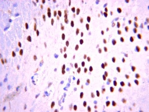
Evaluated by Immunohistochemistry (Paraffin) in Mouse brain tissue sections.Immunohistochemistry (Paraffin) Analysis: A 1:400 dilution of this antibody detected Tbr1 in Mouse cerebral cortex and cerebellum tissue sections.


Tested Applications

Western Blotting Analysis: A 1:500 dilution from a representative lot detected Tbr1 in Mouse fetal brain tissue lysate.Note: Actual optimal working dilutions must be determined by end user as specimens, and experimental conditions may vary with the end user.

Quality

Evaluated by Immunohistochemistry in mouse frontal cortex tissue.

Immunohistochemistry Analysis: 1:400 dilution of this antibody detected Tbr1 in mouse frontal cortex tissue.

Target description

~76 kDa observed; 73.94 kDa calculated. Uncharacterized bands may be observed in some lysate(s).

Physical form

Affinity purified
Purified rabbit polyclonal antibody in buffer containing 0.1 M Tris-Glycine (pH 7.4), 150 mM NaCl with 0.05% sodium azide.

Storage and Stability

Recommended storage: +2°C to +8°C.

Analysis Note

Control
Mouse frontal cortex tissue

Other Notes

Concentration: Please refer to the Certificate of Analysis for the lot-specific concentration.

Disclaimer

Unless otherwise stated in our catalog or other company documentation accompanying the product(s), our products are intended for research use only and are not to be used for any other purpose, which includes but is not limited to, unauthorized commercial uses, in vitro diagnostic uses, ex vivo or in vivo therapeutic uses or any type of consumption or application to humans or animals.

Not finding the right product?  

Try our Product Selector Tool.

Storage Class Code

12 - Non Combustible Liquids

WGK

WGK 1

Flash Point(F)

Not applicable

Flash Point(C)

Not applicable


Certificates of Analysis (COA)

Search for Certificates of Analysis (COA) by entering the products Lot/Batch Number. Lot and Batch Numbers can be found on a product’s label following the words ‘Lot’ or ‘Batch’.

Already Own This Product?

Find documentation for the products that you have recently purchased in the Document Library.

Visit the Document Library

Xue Li et al.
Cerebral cortex (New York, N.Y. : 1991), 30(7), 3960-3976 (2020-02-03)
De novo microdeletion of chromosome 2p15-16.1 presents clinically recognizable phenotypes that include mental retardation, autism, and microcephaly. Chromosomal maintenance 1 (CRM1) is a gene commonly missing in patients with 2p15-16.1 microdeletion and one of two genes found in the smallest
Yuko Muroyama et al.
PLoS genetics, 12(12), e1006514-e1006514 (2016-12-28)
Mitral cells (MCs) of the mammalian olfactory bulb have a single primary dendrite extending into a single glomerulus, where they receive odor information from olfactory sensory neurons (OSNs). Molecular mechanisms for controlling dendritic arbors of MCs, which dynamically change during
Martina Mavrovic et al.
EMBO reports, 21(4), e48880-e48880 (2020-02-18)
KCC2, encoded in humans by the SLC12A5 gene, is a multifunctional neuron-specific protein initially identified as the chloride (Cl- ) extruder critical for hyperpolarizing GABAA receptor currents. Independently of its canonical function as a K-Cl cotransporter, KCC2 regulates the actin
A Docampo-Seara et al.
Brain structure & function, 223(8), 3593-3612 (2018-07-08)
The dorsal part of the developing telencephalon is one of the brain areas that has suffered most drastic changes throughout vertebrate evolution. Its evolutionary increase in complexity was thought to be partly achieved by the appearance of a new neurogenic
Xiao Han et al.
Frontiers in cellular neuroscience, 12, 452-452 (2018-12-12)
The cortex consists of 100s of neuronal subtypes that are organized into distinct functional regions; however, the mechanisms underlying cell fate determination remain unclear. Foxg1 is involved in several developmental processes, including telencephalic patterning, cell proliferation and cell fate determination.

Our team of scientists has experience in all areas of research including Life Science, Material Science, Chemical Synthesis, Chromatography, Analytical and many others.

Contact Technical Service