Skip to Content
MilliporeSigma
All Photos(3)

Key Documents

SAB4300115

Sigma-Aldrich

Anti-phospho-CFL1 (pSer3) antibody produced in rabbit

affinity isolated antibody

Synonym(s):

Anti-CFL antibody produced in rabbit, Anti-cofilin 1 (non-muscle) antibody produced in rabbit

Sign Into View Organizational & Contract Pricing


About This Item

UNSPSC Code:
12352203
NACRES:
NA.41

biological source

rabbit

Quality Level

conjugate

unconjugated

antibody form

affinity isolated antibody

antibody product type

primary antibodies

clone

polyclonal

form

buffered aqueous solution

mol wt

~19 kDa

species reactivity

rat, human, mouse

concentration

1 mg/mL

technique(s)

immunohistochemistry (formalin-fixed, paraffin-embedded sections): 1:50-1:100
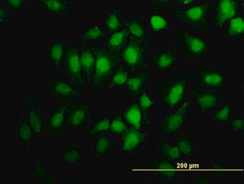
indirect immunofluorescence: 1:100-1:200
western blot: 1:500-1:1000

isotype

IgG

immunogen sequence

(M-A-SP-G-V)

NCBI accession no.

UniProt accession no.

shipped in

wet ice

storage temp.

−20°C

target post-translational modification

phosphorylation (pSer3)

Gene Information

human ... CFL1(1072)

General description

The cofilin 1 (CFL1) gene is mapped to human chromosome 11q13.1. It has four exons. The CFL1 gene codes for a ubiquitously expressed, 18.5 kDa phosphoprotein.

Immunogen

Peptide sequence around phosphorylation site of serine 3 (M-A-S(p)-G-V), according to the protein CFL1.

Application

Anti-phospho-CFL1 (pSer3) antibody produced in rabbit is used in Western Blotting.

Biochem/physiol Actions

Cofilin-l (CFL1) plays a vital role in the regulation of actin cytoskeleton reorganization. It facilitates cytokinesis, cell motility and morphogenesis. CFL1 is also implicated in cancer cell migration and invasion in several types of tumor tissues. Elevated expression of the protein is associated with the development of esophageal squamous cell carcinoma (ESCC). In addition, its abnormal expression leads to vulvar cancer and thus, it can be considered as a potential therapeutic target for the same. Activated CFL1 helps in preventing cytoskeletal-associated defects in axonal development stimulated by synucleins (Syns).

Features and Benefits

Evaluate our antibodies with complete peace of mind. If the antibody does not perform in your application, we will issue a full credit or replacement antibody. Learn more.

Target description

Controls reversibly actin polymerization and depolymerization in a pH-sensitive manner. It has the ability to bind G- and F-actin in a 1:1 ratio of cofilin to actin. It is the major component of intranuclear and cytoplasmic actin rods.

Physical form

Solution in phosphate-buffered saline containing 0.02% sodium azide and 50% glycerol

Disclaimer

Unless otherwise stated in our catalog or other company documentation accompanying the product(s), our products are intended for research use only and are not to be used for any other purpose, which includes but is not limited to, unauthorized commercial uses, in vitro diagnostic uses, ex vivo or in vivo therapeutic uses or any type of consumption or application to humans or animals.

Not finding the right product?  

Try our Product Selector Tool.

Storage Class Code

10 - Combustible liquids

WGK

WGK 1

Flash Point(F)

Not applicable

Flash Point(C)

Not applicable


Certificates of Analysis (COA)

Search for Certificates of Analysis (COA) by entering the products Lot/Batch Number. Lot and Batch Numbers can be found on a product’s label following the words ‘Lot’ or ‘Batch’.

Already Own This Product?

Find documentation for the products that you have recently purchased in the Document Library.

Visit the Document Library

The role of cofilin-l in vulvar squamous cell carcinoma: A marker of carcinogenesis, progression and targeted therapy.
Wu Q, et al.
Oncology Reports, 35(5), 2743-2754 (2016)
Cofilin 1 activation prevents the defects in axon elongation and guidance induced by extracellular alpha-synuclein.
Tilve S, et al.
Scientific Reports, 5, 16524-16524 (2015)
Charnese Bowes et al.
The Journal of cell biology, 218(10), 3258-3271 (2019-09-01)
Actin dynamics is central for cells, and especially for the fast-moving leukocytes. The severing of actin filaments is mainly achieved by cofilin, assisted by Aip1/Wdr1 and coronins. We found that in Wdr1-deficient zebrafish embryos, neutrophils display F-actin cytoplasmic aggregates and
Association between CFL1 gene polymorphisms and spina bifida risk in a California population.
Zhu H, et al.
BMC Medical Genetics, 8(1), 12-12 (2007)
Zhiyong Liu et al.
Oncotarget, 7(42), 68303-68313 (2016-09-15)
CD74, also known as Ii, was initially considered to participate primarily in antigen presentation. Subsequent studies have shown that CD74 is highly expressed in various types of tumor cells and has multiple roles in a variety of biological processes. CD74

Our team of scientists has experience in all areas of research including Life Science, Material Science, Chemical Synthesis, Chromatography, Analytical and many others.

Contact Technical Service