Skip to Content
MilliporeSigma
All Photos(4)

Key Documents

MABE939

Sigma-Aldrich

Anti-phospho Histone H3 (Ser10), clone 6G8B7 Antibody

clone 6G8B7, 1 mg/mL, from rat

Synonym(s):

H3S10P, Histone H3 (phospho Ser10), H3 histone, family 3A, H3.3A, H3 histone, family 3B, H3.3B

Sign Into View Organizational & Contract Pricing


About This Item

UNSPSC Code:
12352203
eCl@ss:
32160702
NACRES:
NA.41

biological source

rat

Quality Level

antibody form

purified antibody

antibody product type

primary antibodies

clone

6G8B7, monoclonal

species reactivity

human

concentration

1 mg/mL

technique(s)

ELISA: suitable
immunocytochemistry: suitable
western blot: suitable

isotype

IgG2aκ

NCBI accession no.

UniProt accession no.

shipped in

wet ice

target post-translational modification

phosphorylation (pSer10)

Gene Information

human ... H3C1(8350)

General description

Histone H3 is one of the five main histone proteins involved in the structure of chromatin in eukaryotic cells. Featuring a main globular domain and a long N-terminal tail, H3 is involved with the structure of the nucleosomes of the ′beads on a string′ structure. The N-terminal tail of histone H3 protrudes from the globular nucleosome core and can undergo several different types of epigenetic modifications that influence cellular processes. These modifications include the covalent attachment of methyl or acetyl groups to lysine and arginine amino acids and the phosphorylation of serine or threonine. Histone H3 variants (H3.1, H3.2 and H3.3) have been implicated in the epigenetic memory of cellular state. Genome-wide patterns of H3.3 are dependent on amino acid sequence and change with cellular differentiation at developmentally regulated loci.

Immunogen

KLH-conjugated linear peptide corresponding to human Histone H3 (Ser10).

Application

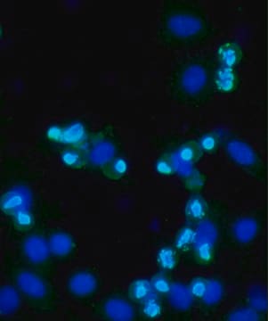
Immunocytochemistry Analysis: A 1:5000 dilution from a representative lot detected Histone H3 (Ser10) in HeLa cells (Prof. Taro Tachibana, Osaka City University).
ELISA Analysis: A representative lot detected Histone H3 (Ser10) using differently phosphorylated and unphosphorylated peptides unmodified H3, H3 S10ph, and H3 S28ph (Prof. Taro Tachibana, Osaka City University).
Research Category
Epigenetics & Nuclear Function
Research Sub Category
Nuclear Receptors
This Anti-phospho Histone H3 (Ser10), clone 6G8B7 Antibody is validated for use in Western Blotting and Immunocytochemistry and ELISA for the detection of phospho Histone H3 (Ser10).

Quality

Evaluated by Western Blotting in HeLa cell lysate.

Western Blotting Analysis: 0.5 µg/mL of this antibody detected Histone H3 (Ser10) in 10 µg of nocodazole treated HeLa cell lysates.

Target description

~17 kDa observed

Linkage

Replaces: 04-1093

Physical form

Format: Purified
Protein G Purified
Purified rat monoclonal IgG2aκ in buffer containing 0.1 M Tris-Glycine (pH 7.4), 150 mM NaCl with 0.05% sodium azide.

Storage and Stability

Stable for 1 year at 2-8°C from date of receipt.

Disclaimer

Unless otherwise stated in our catalog or other company documentation accompanying the product(s), our products are intended for research use only and are not to be used for any other purpose, which includes but is not limited to, unauthorized commercial uses, in vitro diagnostic uses, ex vivo or in vivo therapeutic uses or any type of consumption or application to humans or animals.

Not finding the right product?  

Try our Product Selector Tool.

Storage Class Code

12 - Non Combustible Liquids

WGK

WGK 1

Flash Point(F)

Not applicable

Flash Point(C)

Not applicable


Certificates of Analysis (COA)

Search for Certificates of Analysis (COA) by entering the products Lot/Batch Number. Lot and Batch Numbers can be found on a product’s label following the words ‘Lot’ or ‘Batch’.

Already Own This Product?

Find documentation for the products that you have recently purchased in the Document Library.

Visit the Document Library

Fenghua Bian et al.
Scientific reports, 6, 26061-26061 (2016-05-18)
Uterine stromal cell decidualization is an essential part of the reproductive process. Decidual tissue development requires a highly regulated control of the extracellular tissue remodeling; however the mechanism of this regulation remains unknown. Through systematic expression studies, we detected that
Jian Zhang et al.
Advanced science (Weinheim, Baden-Wurttemberg, Germany), 11(32), e2400719-e2400719 (2024-08-27)
Collective cell migration is a model for nonequilibrium biological dynamics, which is important for morphogenesis, pattern formation, and cancer metastasis. The current understanding of cellular collective dynamics is based primarily on cells moving within a 2D epithelial monolayer. However, solid
Xiaowen Sun et al.
Environmental and molecular mutagenesis, 63(5), 230-245 (2022-06-16)
Genotoxicity testing guidelines require the assessment of the clastogenic and aneugenic potential of compounds. While in vitro micronucleus assays detect both types of endpoints, it requires labor-intensive microscopic scoring and does not discriminate between the two modes of actions. Here
Nuria Cortes-Silva et al.
Current biology : CB, 30(4), 561-572 (2020-02-08)
Accurate chromosome segregation requires assembly of the multiprotein kinetochore complex at centromeres. In most eukaryotes, kinetochore assembly is primed by the histone H3 variant CenH3 (also called CENP-A), which physically interacts with components of the inner kinetochore constitutive centromere-associated network

Our team of scientists has experience in all areas of research including Life Science, Material Science, Chemical Synthesis, Chromatography, Analytical and many others.

Contact Technical Service