Saltar al contenido
Merck

C6805

Sigma-Aldrich

Monoclonal Anti-Collagen, Type VII antibody produced in mouse

clone LH7.2, ascites fluid

Sinónimos:

Anti-EBD1, Anti-EBDCT, Anti-EBR1, Anti-NDNC8

Iniciar sesiónpara Ver la Fijación de precios por contrato y de la organización


About This Item

MDL number:
UNSPSC Code:
12352203
NACRES:
NA.41

biological source

mouse

Quality Level

conjugate

unconjugated

antibody form

ascites fluid

antibody product type

primary antibodies

clone

LH7.2, monoclonal

contains

15 mM sodium azide

species reactivity

human, goat, marmoset, monkey, guinea pig, sheep, pig, bovine

technique(s)

dot blot: suitable
immunocytochemistry: suitable
indirect ELISA: suitable
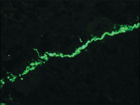
indirect immunofluorescence: 1:1,000 using human or other mammalian frozen sections
western blot: suitable

isotype

IgG1

UniProt accession no.

shipped in

dry ice

storage temp.

−20°C

target post-translational modification

unmodified

Gene Information

human ... COL7A1(1294)

Categorías relacionadas

General description

Collagen is a fibrous protein present in the extracellular framework of all vertebrates. Collagen type VII is the crucial component of anchoring fibrils and mutation in type VII collagen gene (COL7A1) leads to epidermolysis bullosa dystrophica. Monoclonal anti-collagen, type VII antibody can be used to distinguish invasive melanoma from non-invasive melanoma by visualizing the appearance and integrity of epidermal basement membrane. It can also be used in immunoblotting. Mouse anti-collagen type VII antibody reacts specifically with an epitope located on collagenase digested type VII collagen (150 kDa). The product also reacts with the basement membrane zone of stratified squamous epithelia of sheep, pig, bovine, guinea pig, goat and human.
Collagen type VII alpha 1 chain (COL7A1) is majorly expressed in skin, secreted by keratinocytes and fibroblasts. The gene is located on human chromosome 3p21.31.

Immunogen

insoluble fractions of neonatal human foreskin epidermal cells

Application

Monoclonal Anti-Collagen, Type VII antibody produced in mouse has been used in immuno-gold labelling.
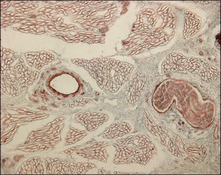
Monoclonal anti-collagen type VII antibody can be used as a primary antibody in virus titration. It can also be used for the localization of type VII collagen in different immunochemical assays like dot blot, immunocytochemistry and ELISA.

Biochem/physiol Actions

Collagen type VII alpha 1 chain (COL7A1) is required for epithelium-to-stroma anchorage in skin, mucosa and cornea. It is considered as an immediate-early response gene for transforming growth factor β (TGF-β)/SMAD (mothers against decapentaplegic) signaling pathway.

Disclaimer

Unless otherwise stated in our catalog or other company documentation accompanying the product(s), our products are intended for research use only and are not to be used for any other purpose, which includes but is not limited to, unauthorized commercial uses, in vitro diagnostic uses, ex vivo or in vivo therapeutic uses or any type of consumption or application to humans or animals.

¿No encuentra el producto adecuado?  

Pruebe nuestro Herramienta de selección de productos.

Optional

Storage Class

12 - Non Combustible Liquids

wgk_germany

nwg

flash_point_f

Not applicable

flash_point_c

Not applicable


Elija entre una de las versiones más recientes:

Certificados de análisis (COA)

Lot/Batch Number

¿No ve la versión correcta?

Si necesita una versión concreta, puede buscar un certificado específico por el número de lote.

¿Ya tiene este producto?

Encuentre la documentación para los productos que ha comprado recientemente en la Biblioteca de documentos.

Visite la Librería de documentos

Daisuke Sawamura et al.
The Journal of investigative dermatology, 118(6), 967-971 (2002-06-13)
The expression of intradermally injected DNA by keratinocytes is found mainly in the upper and middle layers of the epidermis. To investigate the mechanism of this selective expression, we observed the sequential changes in the distribution of interleukin-6-expressing keratinocytes after
COL7A1 editing via CRISPR/Cas9 in recessive dystrophic epidermolysis bullosa
Hainzl S, et al.
Molecular Therapy, 25(11), 2573-2584 (2017)
Arkadii K Beilin et al.
International journal of molecular sciences, 22(4) (2021-03-07)
The recessive form of dystrophic epidermolysis bullosa (RDEB) is a debilitating disease caused by impairments in the junctions of the dermis and the basement membrane of the epidermis. Mutations in the COL7A1 gene induce multiple abnormalities, including chronic inflammation and
SMAD3/4-dependent transcriptional activation of the human type VII collagen gene (COL7A1) promoter by transforming growth factor ?
Vindevoghel L, et al.
Proceedings of the National Academy of Sciences of the USA, 95(25), 14769-14774 (1998)
Martin A Barbier et al.
Current protocols, 2(1), e353-e353 (2022-01-28)
Efficient gene transfer into cultured fibroblasts and keratinocytes during retroviral transduction is a critical step toward the treatment of genodermatoses such as epidermolysis bullosa. However, achieving high transduction rates is still a difficult task, particularly for the insertion of large

Nuestro equipo de científicos tiene experiencia en todas las áreas de investigación: Ciencias de la vida, Ciencia de los materiales, Síntesis química, Cromatografía, Analítica y muchas otras.

Póngase en contacto con el Servicio técnico