Saltar al contenido
Merck
Todas las fotos(2)

Documentos clave

MABF954

Sigma-Aldrich

Anti-LAG3 Antibody, clone 4-10-C9

clone 4-10-C9, from mouse

Sinónimos:

Lymphocyte activation gene 3 protein, CD223, LAG-3

Iniciar sesiónpara Ver la Fijación de precios por contrato y de la organización


About This Item

UNSPSC Code:
12352203
eCl@ss:
32160702
NACRES:
NA.41

biological source

mouse

Quality Level

antibody form

purified immunoglobulin

antibody product type

primary antibodies

clone

4-10-C9, monoclonal

species reactivity

mouse

technique(s)

flow cytometry: suitable
immunocytochemistry: suitable

isotype

IgG2aκ

NCBI accession no.

UniProt accession no.

shipped in

wet ice

target post-translational modification

unmodified

Gene Information

mouse ... Lag3(16768)

General description

Lymphocyte activation gene 3 protein (UniProt Q61790; also known as CD223, LAG-3) is encoded by the Lag3 gene (Gene ID 16768) in murine species. LAG-3 is an inhibitory T-cell surface molecule that modulates T-cell activation and homeostasis. LAG-3 co-localizes with CD8 and CD4 upon TCR engagement and alters TCR signaling. LAG-3 suppresses homeostasis proliferation (HP) of both lymphocytes and some dendritic cell populations in vivo, and enhanced HP is observed in mice deficient in LAG-3. LAG-3 signaling is shown to negatively regulate STAT5 phosphorylation in activated T cells, and no enhancement of HP was seen upon LAG-3 blockade in mice deficient in both STAT5a and STAT5b. Murine LAG-3 is produced with a signal peptide sequence (a.a. 1-22), the removal of which yields the mature protein with a large extracellular region (a.a. 23-442), followed by a transmembrane segment (a.a. 443-463) and a cytoplasmic tail (a.a. 464-521). The extracellular portion contains a V-type Ig-like domain (a.a. 37-163), followed by three C2-type Ig-like domains (a.a. 165-246, 258-341, and 345-412) and elven tandem repeats of 2-amino acid E-X seqeunce at its C-terminal end (a.a. 493-518).

Specificity

Clone 4-10-C9 immunostained surface LAG-3 on activated CD4+ T cells by targeting an extracellular epitope within the third and fourth Ig-like domains (D3/D4 domains; second and third C2-type Ig-like domains). Surface LAG-3 degradation by pronase treatment abolished cell surface staining (Woo, S.R., et al. (2010). Eur. J. Immunol. 40(6):1768-1777).

Immunogen

Epitope: Within D3/D4 domains.
Murine LAG-3-expressing mouse T-cell hybridoma.

Application

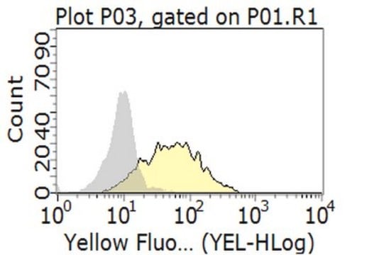
Flow Cytometry Analysis: 2.5 µg/mL from a representative lot detected surface LAG-3 immunoreactivity among the CD4+ and CD8+ populations of wild-type, but not Lag3-knockout, mouse splenocytes activated in vitro via CD3 cross-linking (Courtesy of Dario A. Vignali, Ph.D., University of Pittsburgh, PA, U.S.A.).
Flow Cytometry Analysis: A representative lot was fluorescently conjugated and detected an increased number of LAG-3-positive cells within the CD4+ and CD8+ populations of infiltrating lymphocytes (TILs) in tumors developed in mice exografted with murine B16 melanoma, MC38 colon adenocarcinoma, or Sa1N fibrosarcoma cells (Woo, S.R., et al. (2012). Cancer Res. 72(4): 917–927).
Flow Cytometry Analysis: A representative lot, pre-conjugated with Alexa Fluor 647, detected both surface and intracellular LAG-3 by immunofluorescent staining of non-permeabilized and permeabilized primary murine CD4+ T cells activated in vitro via CD3 & CD28 cross-linking by immobilized antibodies. Pronase treatment of cells prior to permeabilization abolished cell surface staining (Woo, S.R., et al. (2010). Eur. J. Immunol. 40(6):1768-1777).
Flow Cytometry Analysis: A representative lot, pre-conjugated with Alexa Fluor 647, detected a time-dependent recovery of cell surface LAG-3 immunoreactivity on activated murine CD4+ T cells after initial surface LAG-3 degradation by pronase treatment. Protein synthesis inhibitor cycloheximide (Cat. No. 239764) or protein transport inhibitor Brefeldin A (Cat. No. 203729) treatment partially blocked the recovery (Woo, S.R., et al. (2010). Eur. J. Immunol. 40(6):1768-1777).
Immunocytochemistry Analysis: A representative lot detected both surface and intracellular LAG-3 by fluorescent immunocytochemistry staining of non-permeabilized and permeabilized primary murine CD4+ T cells activated in vitro via CD3 & CD28 cross-linking by immobilized antibodies. Pronase treatment of cells prior to permeabilization abolished cell surface staining (Woo, S.R., et al. (2010). Eur. J. Immunol. 40(6):1768-1777).
Immunocytochemistry Analysis: A representative lot detected intracellular LAG-3 immunoreactivity co-localized with those of the early and recycling endosome marker EEA1, as well as endosomal markers Rab11b and Rab27a by fluorescent immunocytochemistry staining of activated murine CD4+ T cells following pronase treatment and permeabilization (Woo, S.R., et al. (2010). Eur. J. Immunol. 40(6):1768-1777).
This Anti-LAG3 Antibody, clone 4-10-C9 is validated for use in Flow Cytometry, Immunocytochemistry for the detection of LAG3.

Quality

Evaluated by Flow Cytometry in mouse splenocytes.

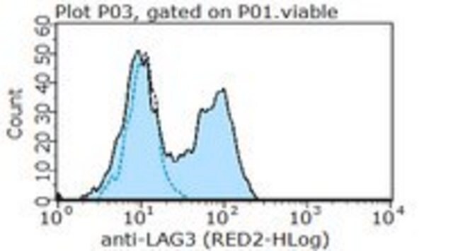
Flow Cytometry Analysis: 1 µg/mL of this antibody detected an induction of LAG-3-positive population in isolated mouse splenocytes following a 3-day 2 µg/mL Concanavalin A (Con A) stimulation.

Target description

54.51/56.98 kDa (mature/pro-form) calculated.

Physical form

Format: Purified

Legal Information

ALEXA FLUOR is a trademark of Life Technologies

¿No encuentra el producto adecuado?  

Pruebe nuestro Herramienta de selección de productos.

Storage Class

12 - Non Combustible Liquids

wgk_germany

WGK 1


Certificados de análisis (COA)

Busque Certificados de análisis (COA) introduciendo el número de lote del producto. Los números de lote se encuentran en la etiqueta del producto después de las palabras «Lot» o «Batch»

¿Ya tiene este producto?

Encuentre la documentación para los productos que ha comprado recientemente en la Biblioteca de documentos.

Visite la Librería de documentos

Timo Eninger et al.
Proceedings of the National Academy of Sciences of the United States of America, 119(24), e2119804119-e2119804119 (2022-06-07)
Single-cell transcriptomics has revealed specific glial activation states associated with the pathogenesis of neurodegenerative diseases, such as Alzheimer’s and Parkinson’s disease. While these findings may eventually lead to new therapeutic opportunities, little is known about how these glial responses are
Yuta Morisaki et al.
Frontiers in cellular neuroscience, 17, 1308972-1308972 (2023-11-29)
Microglia are resident innate immune cells in the central nervous system (CNS) and play important roles in the development of CNS homeostasis. Excessive activation and neurotoxicity of microglia are observed in several CNS disorders, but the mechanisms regulating their activation
Marc Emmenegger et al.
EMBO molecular medicine, 13(9), e14745-e14745 (2021-07-27)
While the initial pathology of Parkinson's disease and other α-synucleinopathies is often confined to circumscribed brain regions, it can spread and progressively affect adjacent and distant brain locales. This process may be controlled by cellular receptors of α-synuclein fibrils, one
Emily Wingrove et al.
Cell reports, 27(4), 1277-1292 (2019-04-25)
The brain is a major site of relapse for several cancers, yet deciphering the mechanisms of brain metastasis remains a challenge because of the complexity of the brain tumor microenvironment (TME). To define the molecular landscape of brain metastasis from

Nuestro equipo de científicos tiene experiencia en todas las áreas de investigación: Ciencias de la vida, Ciencia de los materiales, Síntesis química, Cromatografía, Analítica y muchas otras.

Póngase en contacto con el Servicio técnico