MABF245
Anti-TIM-1 Antibody, clone 3B3
clone 3B3, from rat
Sinónimos:
Hepatitis A virus cellular receptor 1 homolog, HAVcr-1, Kidney injury molecule 1, KIM-1, T cell immunoglobulin and mucin domain-containing protein 1, T-cell immunoglobulin mucin receptor 1, T cell membrane protein 1, TIM-1, TIMD-1
About This Item
Productos recomendados
biological source
rat
Quality Level
antibody form
purified antibody
antibody product type
primary antibodies
clone
3B3, monoclonal
species reactivity
mouse
technique(s)
flow cytometry: suitable
isotype
IgG2aκ
NCBI accession no.
UniProt accession no.
shipped in
ambient
target post-translational modification
unmodified
Gene Information
human ... HAVCR1(26762)
General description
Specificity
Immunogen
Application
Affects Function: A representative lot induced a rapid capping of anti-CD3 bound CD3 on the surface of CD4+ T cells with a concomitant T cell morphology change and increased motility (Xiao, S., et al. (2007). J. Exp. Med. 204(7):1691-1702).
Affects Function: A representative lot enhanced the severity of experimental autoimmune encephalomyelitis (EAE) in mice, while clone RMT1-10 prevented EAE (Xiao, S., et al. (2007). J. Exp. Med. 204(7):1691-1702).
Affects Function: A representative lot enhanced T cell proliferation by crosslinking TIM-1 in vitro. When injected in mice in vivo at the beginning and during OVA sensitization, clone 3B3 prevented tolerance induction and enhanced response of isolated spleen T cells to OVA stimulation in culture (Umetsu, S.E., et al. (2005). Nat. Immunol.6(5):447-454).
Affinity Binding Assay: A representative lot bound TIM-1 with a similar association rate as clone RMT1-10 (Cat. No. MABF225), while clone 3B3 displayed a 10-times slower dissociation rate than clone RMT1-10 (Xiao, S., et al. (2007). J. Exp. Med. 204(7):1691-1702).
ELISA Analysis: Representative lots detected recombinant murine TIM-1, but not TIM-3 or TIM-4, Ig fusion protein. Clone 3B3 displayed an enhanced affinity toward TIM-4 fusion construct lacking the mucin domain when compared to full-length TIM-4 fusion construct (Xiao, S., et al. (2007). J. Exp. Med. 204(7):1691-1702; Umetsu, S.E., et al. (2005). Nat. Immunol.6(5):447-454).
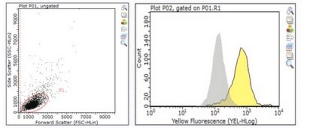
Flow Cytometry Analysis: A representative lot immunostained the surface of TIM-1-transfected, but not untransfected or TIM-2-transfected 300.19 mouse pre-B-cells. A time-dependent increase of CD4+ cell surface TIM-1 expression was detected following TCR activation by APCs or by anti-CD3 and anti-CD28 antibodies (Umetsu, S.E., et al. (2005). Nat. Immunol.6(5):447-454).
Inflammation & Immunology
Quality
Flow Cytometry Analysis: 0.5 µg of this antibody detected the exogenously expressed murine TIM-1 on the surface of one million transfected 300.19 mouse pre-B-cells.
Target description
Physical form
Storage and Stability
Handling Recommendations: Upon receipt and prior to removing the cap, centrifuge the vial and gently mix the solution. Aliquot into microcentrifuge tubes and store at -20°C. Avoid repeated freeze/thaw cycles, which may damage IgG and affect product performance.
Other Notes
Disclaimer
¿No encuentra el producto adecuado?
Pruebe nuestro Herramienta de selección de productos.
Storage Class
12 - Non Combustible Liquids
wgk_germany
WGK 2
flash_point_f
Not applicable
flash_point_c
Not applicable
Certificados de análisis (COA)
Busque Certificados de análisis (COA) introduciendo el número de lote del producto. Los números de lote se encuentran en la etiqueta del producto después de las palabras «Lot» o «Batch»
¿Ya tiene este producto?
Encuentre la documentación para los productos que ha comprado recientemente en la Biblioteca de documentos.
Nuestro equipo de científicos tiene experiencia en todas las áreas de investigación: Ciencias de la vida, Ciencia de los materiales, Síntesis química, Cromatografía, Analítica y muchas otras.
Póngase en contacto con el Servicio técnico


