Skip to Content
Merck
All Photos(2)

Documents

MAB351

Sigma-Aldrich

Anti-Glutamate Decarboxylase Antibody, 65 kDa isoform, clone GAD-6

clone GAD-6, Chemicon®, from mouse

Synonym(s):

GAD65

Sign Into View Organizational & Contract Pricing


About This Item

UNSPSC Code:
12352203
eCl@ss:
32160702
NACRES:
NA.41

biological source

mouse

Quality Level

antibody form

purified immunoglobulin

antibody product type

primary antibodies

clone

GAD-6, monoclonal

species reactivity

rat, human

manufacturer/tradename

Chemicon®

technique(s)

immunohistochemistry: suitable
western blot: suitable

isotype

IgG2a

NCBI accession no.

UniProt accession no.

shipped in

dry ice

target post-translational modification

unmodified

Gene Information

human ... GAD2(2572)

General description

Gutamic acid decarboxylase (GAD; E.C. 4.1.1.15) is the enzyme responsible for the conversion of glutamic acid to gamma-aminobutyric acid (GABA), the major inhibitory transmitter in higher brain regions, and putative paracrine hormone in pancreatic islets. Two molecular forms of GAD (65 kDa and 67 kDa, 64% aa identity between forms) are highly conserved and both forms are expressed in the CNS, pancreatic islet cells, testis, oviduct and ovary. The isoforms are regionally distributed cytoplasmically in the brains of rats and mice (Sheikh, S. et al. 1999). GAD65 is an ampiphilic, membrane-anchored protein (585 a.a.), encoded on human chromosome 10, and is responsible for vesicular GABA production. GAD67 is cytoplasmic (594 a.a.), encoded on chromosome 2, and seems to be responsible for significant cytoplasmic GABA production. GAD expression changes during neural development in rat spinal cord. GAD65 is expressed transiently in commissural axons around E13 but is down regulated the next day while GAD67 expression increases mostly in the somata of those neurons (Phelps, P. et al. 1999). In mature rat pancreas, GAD65 and GAD67 appear to be differentially localized, GAD65 primarily in insulin-containing beta cells and GAD67 in glucagon-containing (A) cells (Li, L. et al. 1995). GAD67 expression seems to be particularly plastic and can change in response to experimental manipulation (for example neuronal stimulation or transection) or disease progression and emergent disorders like schizophrenia (Volk et al., 2000). Colocalization of the two GAD isoforms also shows changes in GAD65/GAD67 distributions correlated with certain disease states such as IDDM and SMS.

Specificity

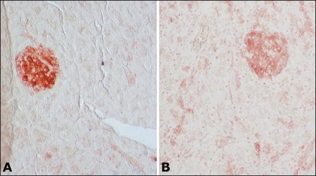
Recognizes the lower molecular weight isoform of the two GAD isoforms identified in brain (Gottlieb, et al., 1986; Chang & Gottlieb, 1988). This monclonal antibody can be used for immunohistochemical localization in brain or pancreas. Anti-GAD has also been used to label purified GAD on Western blots (Chang & Gottlieb, 1988).

Immunogen

Purified rat brain glutamic acid decarboxylase.

Application

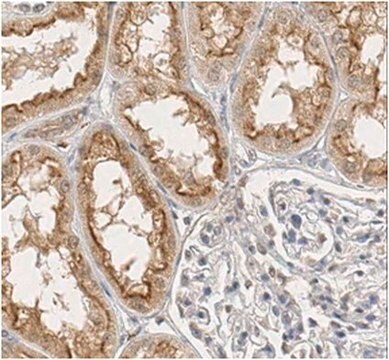
Anti-GAD Antibody. 65 kDa isoform, clone GAD-6 detects level of Glutamate Decarboxylase & has been published & validated for use in IH & WB.
Immunohistochemistry: ( 1 μg/mL * See protocol)

Western blot

Optimal working dilutions must be determined by end user.

APPLICATION NOTES FOR MAB351

IMMUNOHISTOCHEMISTRY

1) Perfuse rats with 100 mM phosphate buffer, pH 7.4 containing 1% paraformaldehyde, 0.34% L-lysine and 0.05% m-periodate (1% PLP).

2) Postfix brains in 1% PLP for 1-2 hours.

3) Transfer brains to 100 mM phosphate buffer containing 30% sucrose and gently agitate on a shaker platform at +4°C for 48-60 hours.

4) Using a sliding microtome, cut 30 mm sections of frozen cerebellum. As the sections are cut, collect them in a vial of cold 100 mM phosphate buffer.

5) Incubate sections in PBS containing 1.5% normal serum and 0.2% Triton X-100 for 30 minutes.

6) On a shaker platform, incubate sections with MAB351 (diluted 1 μg/mL in PBS containing 1.5% normal serum and 0.2% Triton X-100) for 12-36 hours at +4°C.

7) On a shaker platform, rinse sections eight times, 10-15 minutes per rinse, in PBS.

8) Detect with standard secondary antibody detection system (PAP, ABC, etc.).

9) Mount sections, dehydrate, and apply coverslips.
Research Category
Neuroscience
Research Sub Category
Neurotransmitters & Receptors

Target description

65 kDa

Physical form

Format: Purified
Protein A purified
Purified immunoglobulin. Lyophilized from 10 mM potassium phosphate, 70 mM sodium chloride, pH 7.4, 0.02% sodium azide. Reconstitute with 100 μL of sterile water (1 mg/mL).

Storage and Stability

Maintain lyophilized material at -20°C for up to 12 months. After reconstitution maintain frozen at -20°C in undiluted aliquots for up to 6 months. Avoid repeated freeze/thaw cycles.

Analysis Note

Control
Rat brain tissue

human brain lysate

Other Notes

Concentration: Please refer to the Certificate of Analysis for the lot-specific concentration.

Legal Information

CHEMICON is a registered trademark of Merck KGaA, Darmstadt, Germany

Disclaimer

Unless otherwise stated in our catalog or other company documentation accompanying the product(s), our products are intended for research use only and are not to be used for any other purpose, which includes but is not limited to, unauthorized commercial uses, in vitro diagnostic uses, ex vivo or in vivo therapeutic uses or any type of consumption or application to humans or animals.

Not finding the right product?  

Try our Product Selector Tool.

Pictograms

Exclamation mark

Signal Word

Warning

Hazard Statements

Hazard Classifications

Acute Tox. 4 Dermal - Acute Tox. 4 Inhalation - Acute Tox. 4 Oral - Aquatic Chronic 3

Storage Class Code

13 - Non Combustible Solids

WGK

WGK 3

Flash Point(F)

Not applicable

Flash Point(C)

Not applicable


Certificates of Analysis (COA)

Search for Certificates of Analysis (COA) by entering the products Lot/Batch Number. Lot and Batch Numbers can be found on a product’s label following the words ‘Lot’ or ‘Batch’.

Already Own This Product?

Find documentation for the products that you have recently purchased in the Document Library.

Visit the Document Library

Chemical phenotypes of P2X2 purinoreceptor immunoreactive cell bodies in the area postrema.
Mangano, C; Collden, G; Meister, B
Purinergic Signaling null
Richard Fairless et al.
The Journal of neuroscience : the official journal of the Society for Neuroscience, 28(48), 12969-12981 (2008-11-28)
Two families of cell-adhesion molecules, predominantly presynaptic neurexins and postsynaptic neuroligins, are important for the formation and functioning of synapses in the brain, and mutations in several genes encoding these transmembrane proteins have been found in autism patients. However, very
Differential distribution of diacylglycerol lipase-alpha and N-acylphosphatidylethanolamine-specific phospholipase d immunoreactivity in the superficial spinal dorsal horn of rats.
Hegyi, Z; Hollo, K; Kis, G; Mackie, K; Antal, M
Glia null
A Transgenic Mouse Line Expressing the Red Fluorescent Protein tdTomato in GABAergic Neurons.
Besser, S; Sicker, M; Marx, G; Winkler, U; Eulenburg, V; Hulsmann, S; Hirrlinger, J
Testing null
Pei-Rung Wu et al.
Cerebral cortex (New York, N.Y. : 1991), 27(9), 4303-4313 (2016-08-09)
Prenatally, the cytokine CXCL12 regulates cortical interneuron migration, whereas its postnatal functions are poorly understood. Here, we report that CXCL12 is expressed postnatally in layer V pyramidal neurons and localizes on their cell bodies in the medial prefrontal cortex (mPFC)

Our team of scientists has experience in all areas of research including Life Science, Material Science, Chemical Synthesis, Chromatography, Analytical and many others.

Contact Technical Service