Skip to Content
Merck
All Photos(1)

Key Documents

304M-1

Sigma-Aldrich

Nerve Growth Factor Receptor (NGFR) (MRQ-21) Mouse Monoclonal Antibody

Sign Into View Organizational & Contract Pricing


About This Item

UNSPSC Code:
12352203
NACRES:
NA.41

biological source

mouse

Quality Level

100
500

conjugate

unconjugated

antibody form

diluted ascites fluid

antibody product type

primary antibodies

clone

MRQ-21, monoclonal

description

For In Vitro Diagnostic Use in Select Regions (See Chart)

form

buffered aqueous solution

species reactivity

human

packaging

vial of 0.1 mL concentrate (304M-14)
vial of 0.5 mL concentrate (304M-15)
bottle of 1.0 mL predilute (304M-17)
vial of 1.0 mL concentrate (304M-16)
bottle of 7.0 mL predilute (304M-18)

manufacturer/tradename

Cell Marque

technique(s)

immunohistochemistry (formalin-fixed, paraffin-embedded sections): 1:100-1:500

isotype

IgG1

control

breast

shipped in

wet ice

storage temp.

2-8°C

visualization

cytoplasmic

Gene Information

human ... NGFR(4804)

General description

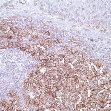
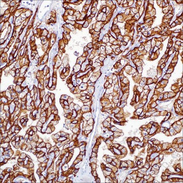
NGFR, a 75 kd glycoprotein (also known as P-75NTR), is the first of neurotrophin receptors to be isolated and is a member of the tumor necrosis factor (TNF) receptor family. It is expressed not only in sympathetic and sensory neurons, but also in various neural crest cell or tumor derivatives such as melanocytes, melanomas, neuroblastomas, pheochromocytomas, neurofibromas, and neurotized nevi (type C melanocytes). NGFR has been shown to be a reliable marker for desmoplastic and neurotropic melanoma by several groups. Although the sensitivity of NGFR staining in desmoplastic melanomas has not been systematically analyzed, all reported cases stain with NGFR. A review of 9 cases of desmoplastic melanomas diagnosed in the Dermatopathology Section of the Department of Dermatology at Boston University School of Medicine between 2001 and 2004 disclosed that all 9 cases of desmoplastic melanomas tested stained positively with NGFR. These findings are in contrast to a study reported by Huttenbach et al in which only 33% of their desmoplastic melanomas stained with NGFR. This difference emphasizes the importance of the choice of monoclonal antibody to obtain consistent results. This staining property of desmoplastic melanoma cells can be useful in diagnosing challenging cases, namely early lesions, suspected cases with negative or weak S-100 staining, and those that must be distinguished from a scar. It is now apparent that expression of NGFR is ubiquitous and not limited to the nervous system, being expressed in mature nonneural cells such as perivascular cells, dental pulp cells, lymphoidal follicular dendritic cells, basal epithelium of oral mucosa and hair follicles, prostate basal cells and myoepithelial cells. Unlike the high-affinity nerve growth factor tyrosine kinase receptors (TrkA, TrkB and TrkC), NGFR has no intrinsic tyrosine kinase activity. Studies in prostate and urothelial cancer suggest that NGFR may act as a tumour suppressor, negatively regulating cell growth and proliferation. Anti-NGFR labels the myoepithelial cells of breast ducts and intralobular fibroblasts of breast ducts and thus aides in the diagnosis of malignancy in the breast. Associated products: GFAP, S.M. Myosin, Calponin, NSE, Myelin Basic Protein, S-100, HMB45, Tyrosinase, MART-1

Quality


IVD

IVD

IVD

RUO

Linkage

Nerve Growth Factor Receptor (NGFR) Positive Control Slides, Product No. 304S, are available for immunohistochemistry (formalin-fixed, paraffin-embedded sections).

Physical form

Solution in Tris Buffer, pH 7.3-7.7, with 1% BSA and <0.1% Sodium Azide

Preparation Note

Download the IFU specific to your product lot and formatNote: This requires a keycode which can be found on your packaging or product label.

Other Notes

For Technical Service please contact: 800-665-7284 or email: service@cellmarque.com

Legal Information

Cell Marque is a trademark of Merck KGaA, Darmstadt, Germany

Not finding the right product?  

Try our Product Selector Tool.


Choose from one of the most recent versions:

Certificates of Analysis (COA)

Lot/Batch Number

Don't see the Right Version?

If you require a particular version, you can look up a specific certificate by the Lot or Batch number.

Already Own This Product?

Find documentation for the products that you have recently purchased in the Document Library.

Visit the Document Library

W B Laskin et al.
Human pathology, 31(10), 1230-1241 (2000-11-09)
In contrast with the myxoid variant of neurothekeoma (nerve sheath myxoma), evidence of neurosustentacular (NS) differentiation in the so-called cellular and mixed (intermediate) variants of neurothekeoma remains controversial. In this study, we selected 22 tumors coded as neurothekeoma or nerve
Y Liang et al.
Journal of cutaneous pathology, 25(4), 189-198 (1998-06-03)
The mast cell, which is a histamine-containing cell, has been found to have far more functions in skin inflammation than hitherto understood. To investigate the appearance of mast cells in prurigo nodularis, histamine immunohistochemistry in combination with nerve growth factor
Rebecca Lewis Kelso et al.
Dermatologic surgery : official publication for American Society for Dermatologic Surgery [et al.], 32(2), 177-183 (2006-01-31)
Perineural invasion (PNI) in cutaneous squamous cell carcinoma (CSCC) may portend a poor prognosis for patients. p75NGFR (nerve growth factor receptor) is part of a membrane receptor complex that binds nerve growth factor. Its use for detecting PNI in CSCC
Y Liang et al.
The Journal of investigative dermatology, 111(1), 114-118 (1998-07-17)
The nerve growth factor and its receptor are important in nerve growth, differentiation, maturation, and maintenance. In order to explore the exact distribution of p75 low-affinity nerve growth factor receptor (p75 NGFr) expression in cutaneous nerve fibers, p75 NGFr and
Y Liang et al.
Archives of dermatological research, 291(1), 14-21 (1999-02-20)
Prurigo nodularis is an inflammatory skin disease characterized by neurohyperplasia. Neurotrophins and their receptors play a critical role in nerve growth, differentiation, maturation and maintenance, including cutaneous nerve fiber growth and innervation. They may also be responsible for events related

Articles

IHC antibodies enhance dermatopathology beyond H&E stained slides, improving techniques and applications for dermatological research.

IHC antibodies enhance dermatopathology beyond H&E stained slides, improving techniques and applications for dermatological research.

IHC antibodies enhance dermatopathology beyond H&E stained slides, improving techniques and applications for dermatological research.

IHC antibodies enhance dermatopathology beyond H&E stained slides, improving techniques and applications for dermatological research.

Our team of scientists has experience in all areas of research including Life Science, Material Science, Chemical Synthesis, Chromatography, Analytical and many others.

Contact Technical Service