Z724300
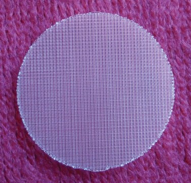
3D Biotek 3D Insert™ PS scaffold
for 96 well plates, 24 inserts/96 well plate
Synonym(s):
3D, 3D Cell Culture, 3D scaffolds, Scaffolds, cell scaffolds
About This Item
clear wells
polystyrene
Recommended Products
material
clear polystyrene
clear wells
polystyrene
description
polystyrene (PS)
sterility
sterile
quality
24 inserts/96 well plate
packaging
pack of 24 ea (24 inserts supplied in 96 well plate)
manufacturer/tradename
3D Biotek PS152096-24
size
96 wells
pore size
~200 μm (Fiber spacing)
binding type
non-treated surface
Looking for similar products? Visit Product Comparison Guide
General description
- Terminally sterilized by gamma irradiation and ready for use
- Compatible with most of your current 2D assays
- Easy for imaging. Cell growth can be monitored by light microscope, no need to purchase any additional sophisticated equipment.
- 96 well compatible
- Fiber Diameter: 150u
- Pore Size: 200u
- 24 Inserts/Pack
Legal Information
Choose from one of the most recent versions:
Certificates of Analysis (COA)
Sorry, we don't have COAs for this product available online at this time.
If you need assistance, please contact Customer Support.
Already Own This Product?
Find documentation for the products that you have recently purchased in the Document Library.
Customers Also Viewed
Articles
3D cell culture overview. Learn about 2D vs 3D cell culture, advantages of 3D cell culture, and techniques available to develop 3D cell models
3D cell culture overview. Learn about 2D vs 3D cell culture, advantages of 3D cell culture, and techniques available to develop 3D cell models
3D cell culture overview. Learn about 2D vs 3D cell culture, advantages of 3D cell culture, and techniques available to develop 3D cell models
3D cell culture overview. Learn about 2D vs 3D cell culture, advantages of 3D cell culture, and techniques available to develop 3D cell models
Our team of scientists has experience in all areas of research including Life Science, Material Science, Chemical Synthesis, Chromatography, Analytical and many others.
Contact Technical Service


