T9026
Monoclonal Anti-α-Tubulin antibody produced in mouse

clone DM1A, ascites fluid
Synonym(s):
Monoclonal Anti-α-Tubulin
About This Item
Recommended Products
biological source
mouse
Quality Level
conjugate
unconjugated
antibody form
ascites fluid
antibody product type
primary antibodies
clone
DM1A, monoclonal
mol wt
antigen ~50 kDa
contains
15 mM sodium azide
species reactivity
yeast, mouse, amphibian, human, rat, chicken, fungi, bovine
enhanced validation
independent ( antibodies)
Learn more about Antibody Enhanced Validation
technique(s)
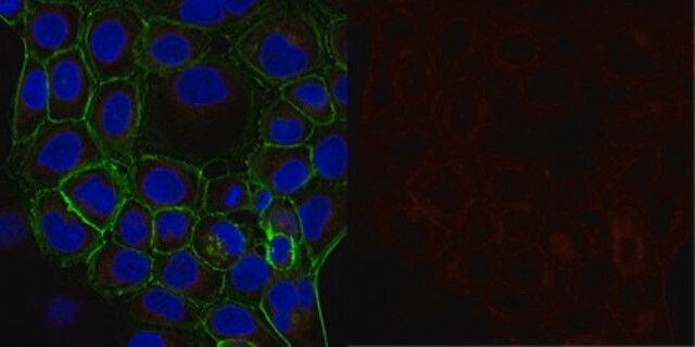
indirect immunofluorescence: 1:500 using cultured chicken fibroblasts
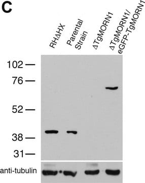
western blot: 1:500 using human or chicken fibroblasts
isotype
IgG1
UniProt accession no.
application(s)
research pathology
shipped in
dry ice
storage temp.
−20°C
target post-translational modification
unmodified
Gene Information
human ... TUBA4A(7277)
mouse ... Tuba1a(22142)
rat ... Tuba1a(64158)
Looking for similar products? Visit Product Comparison Guide
Related Categories
General description
Monoclonal Anti-α-Tubulin (mouse IgG1 isotype) is derived from the DM1A hybridoma produced by the fusion of mouse myeloma cells and splenocytes from immunized BALB/c mice. Purified chick brain microtubules were used as immunogen. The isotype is determined by a double diffusion immunoassay using Mouse Monoclonal Antibody Isotyping Reagents, Product Number ISO2. The product is Protein A purified Monoclonal Anti-α-Tubulin antibody conjugated to fluorescein isothiocyanate, isomer I. It is purified by gel filtration and contains no detectable free FITC. Anti-α-Tubulin FITC antibody, Mouse monoclonal specifically recognizes an epitope in the carboxy terminal part of α-tubulin. It localizes α-tubulin in human, monkey, bovine, chicken, goat, murine, rat, gerbil, hamster, rat kangaroo, amphibia, sea urchin, trypanosome, yeast, fungi and tobacco.
Specificity
Immunogen
Application
Monoclonal Anti-α-Tubulin antibody produced in mouse has been used in the detection of α-Tubulin:
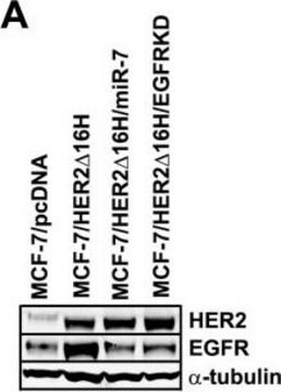
- in human osteosarcoma and in breast cancer cell lines by western blotting
- in HeLa cells by immunofluorescence microscopy
- by immunostaining
- immunohistochemical detection in Xenopus embryos
Biochem/physiol Actions
α/β-Tubulin is the major building block of microtubules. These intracellular, hollow, cylindrical, filamentous structures are present in virtually all eukaryotic cells. Self-assembly of α/β-tubulin leads to polar microtubular structures built from linearly arranged strings of alternatively α- and β-tubulin pairs pointing in the same direction. Microtubules function as structural and mobile elements in mitosis, intracellular transport, ciliary flagellar motility and generation and maintenance of cell shape. α/β-Tubulin and γ-tubulin are members of the tubulin superfamily of proteins. α/β-Tubulin is a heterodimer which consists of one α-tubulin chain and one β-tubulin chain; each subunit has a molecular weight of 55 kDa and they share considerable homolog.
Storage and Stability
For extended storage, the solution may be frozen in working aliquots. Repeated freezing and thawing, or storage in "frost-free" freezers, is not recommended. If slight turbidity occurs upon prolonged storage, clarify the solution by centrifugation before use.
Disclaimer
Not finding the right product?
Try our Product Selector Tool.
Storage Class Code
10 - Combustible liquids
WGK
WGK 3
Flash Point(F)
Not applicable
Flash Point(C)
Not applicable
Choose from one of the most recent versions:
Certificates of Analysis (COA)
Don't see the Right Version?
If you require a particular version, you can look up a specific certificate by the Lot or Batch number.
Already Own This Product?
Find documentation for the products that you have recently purchased in the Document Library.
Customers Also Viewed
Articles
Immunofluorescence uses antibody-conjugated fluorescent molecules for protein localization, modification confirmation, and protein complex visualization.
Immunofluorescence uses antibody-conjugated fluorescent molecules for protein localization, modification confirmation, and protein complex visualization.
Immunofluorescence uses antibody-conjugated fluorescent molecules for protein localization, modification confirmation, and protein complex visualization.
Immunofluorescence uses antibody-conjugated fluorescent molecules for protein localization, modification confirmation, and protein complex visualization.
Our team of scientists has experience in all areas of research including Life Science, Material Science, Chemical Synthesis, Chromatography, Analytical and many others.
Contact Technical Service


