Skip to Content
Merck
All Photos(1)

Key Documents

348M-9

Sigma-Aldrich

WT1 (6F-H2) Mouse Monoclonal Antibody

Sign Into View Organizational & Contract Pricing


About This Item

UNSPSC Code:
12352203
NACRES:
NA.41
clone:
6F-H2, monoclonal
application:
IHC (p)
species reactivity:
human
technique(s):
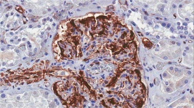
immunohistochemistry (formalin-fixed, paraffin-embedded sections): 1:100-1:500
citations:
14

biological source

mouse

Quality Level

100
500

conjugate

unconjugated

antibody form

culture supernatant

antibody product type

primary antibodies

clone

6F-H2, monoclonal

description

For In Vitro Diagnostic Use in Select Regions (See Chart)

form

buffered aqueous solution

species reactivity

human

packaging

vial of 0.1 mL concentrate (348M-94)
vial of 0.5 mL concentrate (348M-95)
bottle of 1.0 mL predilute (348M-97)
vial of 1.0 mL concentrate (348M-96)
bottle of 7.0 mL predilute (348M-98)

manufacturer/tradename

Cell Marque

technique(s)

immunohistochemistry (formalin-fixed, paraffin-embedded sections): 1:100-1:500

isotype

IgG1κ

control

kidney, mesothelioma, ovarian carcinoma (serous), testis

shipped in

wet ice

storage temp.

2-8°C

visualization

nuclear

Gene Information

human ... WT1(7490)

General description

Wilms’ tumor 1 protein (WT1) is a zinc finger transcription factor, normally expressed intissues of mesodermal origin. The Wilms’ tumor gene encodes a protein that functionsas a tumor suppressor gene mapped to human chromosome 11p13.2 Nuclear WT1immunoreactivity has been observed in high frequencies in several types of mesoderm-derived tumors including epithelioid mesothelioma and ovarian serous carcinoma and thus isused as a marker for these neoplasms.

Quality


IVD

IVD

IVD

RUO

Linkage

WT-1 Positive Control Slides, Product No. 348S, are available for immunohistochemistry (formalin-fixed, paraffin-embedded sections).

Physical form

Solution in Tris Buffer, pH 7.3-7.7, with 1% BSA and <0.1% Sodium Azide

Preparation Note

Download the IFU specific to your product lot and formatNote: This requires a keycode which can be found on your packaging or product label.

Other Notes

For Technical Service please contact: 800-665-7284 or email: service@cellmarque.com

Legal Information

Cell Marque is a trademark of Merck KGaA, Darmstadt, Germany

Not finding the right product?  

Try our Product Selector Tool.


Choose from one of the most recent versions:

Certificates of Analysis (COA)

Lot/Batch Number

Don't see the Right Version?

If you require a particular version, you can look up a specific certificate by the Lot or Batch number.

Already Own This Product?

Find documentation for the products that you have recently purchased in the Document Library.

Visit the Document Library

Alberto M Marchevsky
Archives of pathology & laboratory medicine, 132(3), 397-401 (2008-03-06)
The diagnosis of malignant mesothelioma (MM) is rendered with the aid of immunohistochemistry to demonstrate the presence of "mesothelial," "epithelial," or "sarcomatous" differentiation. Antibody panels that have been proposed for the distinction between MM and other neoplasms usually include 2
Rena J May et al.
Clinical cancer research : an official journal of the American Association for Cancer Research, 13(15 Pt 1), 4547-4555 (2007-08-03)
Wilms' tumor 1 protein (WT1), a transcription factor overexpressed in malignant mesothelioma, leukemias, and other solid tumors, is an ideal target for immunotherapy. WT1 class I peptide epitopes that were identified and shown to stimulate CD8(+) T cells are being
M A Ghanem et al.
Clinical cancer research : an official journal of the American Association for Cancer Research, 6(11), 4265-4271 (2000-12-06)
Wilms' tumor is one of the most common solid tumors of children. The protein product of the tumor-suppressor gene, Wilms' tumor 1 (WT-1), binds to the same DNA sequences as the protein product of the early growth response 1 (EGR-1)
Martin Köbel et al.
International journal of gynecological pathology : official journal of the International Society of Gynecological Pathologists, 35(5), 430-441 (2016-03-15)
There are 5 major histotypes of ovarian carcinomas. Diagnostic typing criteria have evolved over time, and past cohorts may be misclassified by current standards. Our objective was to reclassify the recently assembled Canadian Ovarian Experimental Unified Resource and the Alberta
Sarah K Galfione et al.
International journal of clinical and experimental pathology, 7(5), 2536-2543 (2014-06-27)
Vascular lesions are commonly encountered in routine pathologic practice and often pose diagnostic challenges owing to their morphologic diversity. Although WT-1 expression was reported in some vascular tumors, little is known about its staining patterns in a spectrum of vascular

Our team of scientists has experience in all areas of research including Life Science, Material Science, Chemical Synthesis, Chromatography, Analytical and many others.

Contact Technical Service