Skip to Content
Merck
All Photos(3)

Key Documents

MABT409

Sigma-Aldrich

Anti-Integrin Beta1, clone AIIB2 (Azide Free) Antibody

clone AIIB2, 1 mg/mL, from rat

Synonym(s):

Integrin beta-1, Fibronectin receptor subunit beta, Glycoprotein Iia, GPIIA, VLA-4 subunit beta, CD29

Sign Into View Organizational & Contract Pricing


About This Item

UNSPSC Code:
12352203
eCl@ss:
32160702
NACRES:
NA.41
clone:
AIIB2, monoclonal
application:
ICC
IHC
IP
WB
activity assay
species reactivity:
rhesus macaque, horse, human, pig, canine, bovine, sheep
technique(s):
activity assay: suitable
immunocytochemistry: suitable
immunohistochemistry: suitable
immunoprecipitation (IP): suitable
western blot: suitable
citations:
21

biological source

rat

Quality Level

antibody form

purified antibody

antibody product type

primary antibodies

clone

AIIB2, monoclonal

species reactivity

rhesus macaque, horse, human, pig, canine, bovine, sheep

concentration

1 mg/mL

technique(s)

activity assay: suitable
immunocytochemistry: suitable
immunohistochemistry: suitable
immunoprecipitation (IP): suitable
western blot: suitable

isotype

IgG1κ

NCBI accession no.

UniProt accession no.

shipped in

dry ice

target post-translational modification

unmodified

Gene Information

human ... ITGB1(3688)

General description

Integrin beta-1, also known as Fibronectin receptor subunit beta (FNRB), and Glycoprotein IIa (GPIIA), is a transmembrane glycoprotein that forms noncovalent complexes with various alpha integrin subunits to form the functional receptors that bind to specific extracellular matrix proteins. Integrin receptors are involved in cell adhesion and recognition in a variety of processes including embryogenesis, hemostasis, tissue repair, immune response and metatastatic diffusion of tumor cells. Interactions between integrins and extracellular matrix lead to activation of signal transduction pathways and regulation of gene expression. Phosphorylation of threonines 788 and 789 on integrin beta1 receptor may play a key role in cell-cycle dependent regulation.

Immunogen

Whole cells corresponding to human Integrin Beta1, clone AIIB2 (Azide Free).

Application

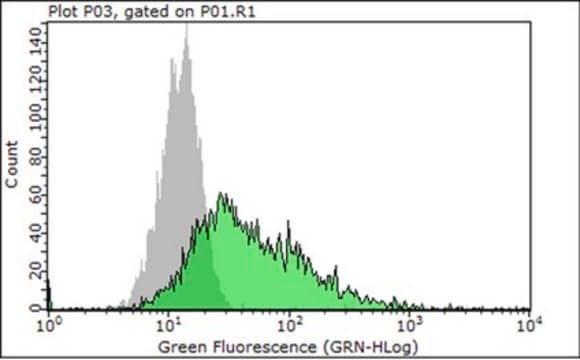
Activity Assay Analysis: A representative lot detected Integrin Beta1, clone AIIB2 (Azide Free) in this functionally blocking antibody abrogated AGS cell attachment to fibronectin while causing only a slight inhibition of cell attachment to vitronectin (Hutton, M., et. al. (2010). Infection and Immunity. 78(11):4523-4531).
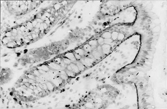
Immunohistochemistry Analysis: A representative lot detected Integrin Beta1, clone AIIB2 (Azide Free) in floating villi tissue (Damsky, C. H., et al. (1992). Journal of Clinical Investigation. 89:210-222).
Immunohistochemistry Analysis: A representative lot detected Integrin Beta1, clone AIIB2 (Azide Free) in CTB aggregates that have invaded matrigel in vitro (Damsky, C. H., et al. (1994). Development. 120:3657-3666).
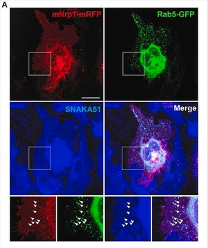
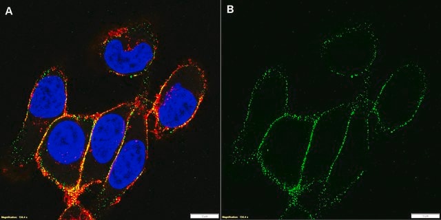
Immunocytochemistry Analysis: A representative lot detected Integrin Beta1, clone AIIB2 (Azide Free) in AGS cells (Hutton, M., et. al. (2010). Infection and Immunity. 78(11):4523-4531).
Immunoprecipitation Analysis: A representative lot immunoprecipitated Integrin Beta1, clone AIIB2 (Azide Free) in NP-40 cell lysate (Werb, Z., et al. (1989). JCB. 109:877-889).
Research Category
Cell Structure
Research Sub Category
Adhesion (CAMs)
This Anti-Integrin Beta1, clone AIIB2 (Azide Free) Antibody is validated for use in Western Blotting and Immunocytochemistry and Activity Assay and Immunoprecipitation and Immunohistochemistry for the detection of Integrin Beta1.

Quality

Evaluated by Western Blotting in HUVEC cell lysate.

Western Blotting Analysis: 1.0 µg/mL of this antibody detected Integrin Beta1, clone AIIB2 (Azide Free) in 10 µg of HUVEC cell lysate.

Target description

~110 kDa observed. The calculated molecular weight of this protein is 88 kDa, but can be observed at ~110 kDa due to high glycosylation.

Physical form

Format: Purified
Protein G Purified
Purified rat monoclonal IgG1κ in buffer containing PBS without preservatives.

Storage and Stability

Stable for 1 year at -20°C from date of receipt.
Handling Recommendations: Upon receipt and prior to removing the cap, centrifuge the vial and gently mix the solution. Aliquot into microcentrifuge tubes and store at -20°C. Avoid repeated freeze/thaw cycles, which may damage IgG and affect product performance.

Disclaimer

Unless otherwise stated in our catalog or other company documentation accompanying the product(s), our products are intended for research use only and are not to be used for any other purpose, which includes but is not limited to, unauthorized commercial uses, in vitro diagnostic uses, ex vivo or in vivo therapeutic uses or any type of consumption or application to humans or animals.

Not finding the right product?  

Try our Product Selector Tool.

Storage Class Code

12 - Non Combustible Liquids

WGK

WGK 2

Flash Point(F)

Not applicable

Flash Point(C)

Not applicable


Certificates of Analysis (COA)

Search for Certificates of Analysis (COA) by entering the products Lot/Batch Number. Lot and Batch Numbers can be found on a product’s label following the words ‘Lot’ or ‘Batch’.

Already Own This Product?

Find documentation for the products that you have recently purchased in the Document Library.

Visit the Document Library

Cecilia Gardelli et al.
Cancer science, 112(1), 217-230 (2020-10-18)
In lung cancer, CD133+ cells represent the subset of cancer stem cells (CSC) able to sustain tumor growth and metastatic dissemination. CSC function is tightly regulated by specialized niches composed of both stromal cells and extracellular matrix (ECM) proteins, mainly
Tsunaki Hongu et al.
Nature cancer, 3(4), 486-504 (2022-04-27)
Disseminated cancer cells frequently lodge near vasculature in secondary organs. However, our understanding of the cellular crosstalk invoked at perivascular sites is still rudimentary. Here, we identify intercellular machinery governing formation of a pro-metastatic vascular niche during breast cancer colonization
Jinhua Zhang et al.
Scientific reports, 7(1), 12099-12099 (2017-09-25)
S100A4 has been implicated in cancer and several inflammatory diseases, but its role in inflammatory bowel disease has not been well investigated. Here, upon infection with Citrobacter rodentium, a model for enteropathogenic Escherichia coli infection in humans, induced the infiltration
Qing Zhao et al.
PLoS pathogens, 14(10), e1007359-e1007359 (2018-10-27)
Translocation of the Helicobacter pylori (Hp) cytotoxin-associated gene A (CagA) effector protein via the cag-Type IV Secretion System (cag-T4SS) into host cells is a hallmark of infection with Hp and a major risk factor for severe gastric diseases, including gastric
Anh Duy Do et al.
Frontiers in immunology, 12, 796177-796177 (2022-02-01)
Helicobacter pylori is a Gram-negative pathogen that can increase the risk of stomach cancer in infected patients. H. pylori exploits lipid rafts to infect host cells. Infection triggers clustering of Lewis x antigen (Lex) and integrins in lipid rafts to

Our team of scientists has experience in all areas of research including Life Science, Material Science, Chemical Synthesis, Chromatography, Analytical and many others.

Contact Technical Service