Skip to Content
Merck
All Photos(1)

Key Documents

MABF1978

Sigma-Aldrich

Anti-Complement C3a/C3a (desArg) Antibody, clone K13/16

clone K13/16, from mouse

Synonym(s):

Complement C3a, C3 and PZP-like alpha-2-macroglobulin domain-containing protein 1

Sign Into View Organizational & Contract Pricing


About This Item

UNSPSC Code:
12352203
eCl@ss:
32160702
NACRES:
NA.41
clone:
K13/16, monoclonal
application:
FACS
IHC
Neutral
species reactivity:
human
technique(s):
flow cytometry: suitable
immunohistochemistry: suitable (paraffin)
neutralization: suitable
citations:
1

biological source

mouse

Quality Level

antibody form

purified antibody

antibody product type

primary antibodies

clone

K13/16, monoclonal

species reactivity

human

packaging

antibody small pack of 25 μL

technique(s)

flow cytometry: suitable
immunohistochemistry: suitable (paraffin)
neutralization: suitable

isotype

IgG1κ

NCBI accession no.

UniProt accession no.

shipped in

ambient

target post-translational modification

unmodified

Gene Information

human ... C3(718)

General description

Complement C3 (UniProt: P01024; also known as C3 and PZP-like alpha-2-macroglobulin domain-containing protein 1) is encoded by the C3 (also known as CPAMD1) gene (Gene ID: 718) in human. C3 is a secreted protein that plays a key role in the activation of the complement system. It s processing by C3 convertase is the central reaction in both classical and alternative complement pathways. C3 has an anaphylatoxon-like domain (aa 693-728) and a NTR (Netrin) domain (aa 1518-1661). IC3 precursor is first processed by the removal of four Arginine residues, forming two chains, beta and alpha, linked by a disulfide bond. C3 convertase activates C3 by cleaving the alpha chain, releasing C3a anaphylatoxin and generating C3b. C3a appears to be important in many inflammatory respons-es and the C3b fragment covalently binds to the cell or bacterial surface and plays a role in opsonisation. C3a anaphylatoxin is a mediator of local inflammatory process. In chronic inflammation, it acts as a chemoattractant for neutrophils. Defects in C3 gene can cause complement component 3 deficiency that leads to recurrent, severe, pyogenic infections because of ineffective opsonization of pathogens. Defects in C3 gene also cause age-related macular degeneration and hemolytic uremic syndrome that can lead to hemolytic anemia and renal failure

Specificity

Clone K13/16 recognizes complement C3a in human tissues. It targets an epitope that is present on human C3, C3a, and C3a-desArg.

Immunogen

A full length native C3a fragment purified from human serum that was activated at 37°C for 1 hour by treatment with 10 mg/mL zymosan.

Application

Anti-Complement C3a/C3a (desArg), clone K13/16, Cat. No. MABF1978, is a mouse monoclonal antibody that detects Complement C3a and has been tested for use in Flow Cytometry, Immunohistochemistry (Paraffin), Neutralization, and Agonist and Inhibition studies.
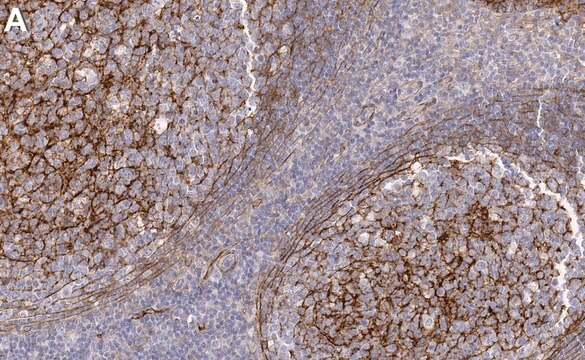
Immunohistochemistry Analysis: A 1:50 dilution from a representative lot detected Complement C3a/C3a (desArg) in human liver and human tonsil tissue.

Agonist or Inhibitor Analysis: Administration of C3a induces a transient influx of Ca2+ release in a dose dependent manner (fluo-3 staining on human PMNs). In this regard, it functions as an agonist for the C3a receptor (Elsner, J., et. al. (1994). Blood. 83(11):3324-31).

Neutralizing Analysis: A representative lot detected Complement C3a/C3a (desArg) in Neutralizing applications (Elsner, J., et. al. (1994). Blood. 83(11):3324-31).

Flow Cytometry Analysis: A representative lot detected Complement C3a/C3a (desArg) in Flow Cytometry applications (Elsner, J., et. al. (1994). Blood. 83(11):3324-31).
Research Category
Inflammation & Immunology

Quality

Evaluated by Immunohistochemistry in human kidney tissue.

Immunohistochemistry Analysis: A 1:50 dilution of this antibody detected Complement C3a/C3a (desArg) in human kidney tissue sections.

Target description

187.15 kDa calculated.

Physical form

Format: Purified
Protein G purified
Purified mouse monoclonal antibody IgG1 in buffer containing 0.1 M Tris-Glycine (pH 7.4), 150 mM NaCl with 0.05% sodium azide.

Storage and Stability

Stable for 1 year at 2-8°C from date of receipt.

Other Notes

Concentration: Please refer to lot specific datasheet.

Disclaimer

Unless otherwise stated in our catalog or other company documentation accompanying the product(s), our products are intended for research use only and are not to be used for any other purpose, which includes but is not limited to, unauthorized commercial uses, in vitro diagnostic uses, ex vivo or in vivo therapeutic uses or any type of consumption or application to humans or animals.

Not finding the right product?  

Try our Product Selector Tool.


Certificates of Analysis (COA)

Search for Certificates of Analysis (COA) by entering the products Lot/Batch Number. Lot and Batch Numbers can be found on a product’s label following the words ‘Lot’ or ‘Batch’.

Already Own This Product?

Find documentation for the products that you have recently purchased in the Document Library.

Visit the Document Library

Maria Jesus Iglesias et al.
Nature communications, 14(1), 3280-3280 (2023-06-08)
Venous thromboembolism (VTE) is a common, multi-causal disease with potentially serious short- and long-term complications. In clinical practice, there is a need for improved plasma biomarker-based tools for VTE diagnosis and risk prediction. Here we show, using proteomics profiling to

Our team of scientists has experience in all areas of research including Life Science, Material Science, Chemical Synthesis, Chromatography, Analytical and many others.

Contact Technical Service