Skip to Content
Merck
All Photos(2)

Key Documents

MAB363

Sigma-Aldrich

Anti-NMDAR1 Antibody, clone 54.1

clone 54.1, Chemicon®, from mouse

Synonym(s):

N-methyl-D-aspartate receptor channel, subunit zeta-1, N-methyl-D-aspartate receptor subunit NR1, NMDA receptor 1, glutamate [NMDA] receptor subunit zeta 1, glutamate receptor, ionotropic, N-methyl D-aspartate 1

Sign Into View Organizational & Contract Pricing


About This Item

UNSPSC Code:
12352203
eCl@ss:
32160702
NACRES:
NA.41
clone:
54.1, monoclonal
application:
ELISA
ICC
IHC (p)
RIA
species reactivity:
rat, monkey, human, Xenopus
technique(s):
ELISA: suitable
immunocytochemistry: suitable
immunohistochemistry (formalin-fixed, paraffin-embedded sections): suitable
radioimmunoassay: suitable
citations:
115

biological source

mouse

Quality Level

antibody form

purified immunoglobulin

antibody product type

primary antibodies

clone

54.1, monoclonal

species reactivity

rat, monkey, human, Xenopus

manufacturer/tradename

Chemicon®

technique(s)

ELISA: suitable
immunocytochemistry: suitable
immunohistochemistry (formalin-fixed, paraffin-embedded sections): suitable
radioimmunoassay: suitable

isotype

IgG2a

NCBI accession no.

UniProt accession no.

shipped in

dry ice

target post-translational modification

unmodified

Gene Information

frog ... Grin1(780048)
human ... GRIN1(2902)
rat ... Grin1(24408)
rhesus monkey ... Grin1(574380)

General description

Glutamate is a widely utilized excitatory neurotransmitter in mammalian brain. The ion channels activated by glutamate are typically divided into two classes. Those that are sensitive to N-methyl-D-aspartate (NMDA) are designated NMDA receptors (NMDAR) while those activated by kainate (KA) and a-amino-3-hydroxy-5-methyl-4-isoxalone propionic acid (AMPA) are referred to as non-NMDA receptors. Two distinct genes encode the NMDA receptor subunits, NR1 and NR2. The NR1 subunit is expressed in abundance in virtually all regions of the brain.

Specificity

Recognizes an epitope between amino acids 660-811 of the NMDAR1 receptor. Shows no cross-reactivity with NMDAR2, NMDAR3, NMDAR4 or NMDAR5.

Immunogen

Epitope: Extracellular loop between transmembrane regions III & IV.
Recombinant fusion protein containing GST and NMDAR1 aa. 660-811.

Application

Anti-NMDAR1 Antibody, clone 54.1 is an antibody against NMDAR1 for use in ELISA, IC, IH, IH(P), RIA.
Immunocytochemistry:
A previous lot of this antibody was used in IC.

RIA:
A previous lot of this antibody was used in RIA.

ELISA:
A previous lot of this antibody was used in ELISA.

Immunohistochemistry: 1:500-2000, on 2-4% paraformaldehyde fixed tissue: typically free-floating, frozen or vibratome sections 24-48 hours at 4°C. Paraffin embedded formalin fixed sections have also been used at similar dilutions and incubation times. Care should be taken not to over fix the tissues. The clone 54.1 has been used for both light and electron microscopy.

Western blot:
1:500 with 50 migrograms/lane of material. Recognizes a 103-116kDa band.

Optimal working dilutions must be determined by end user.
Research Category
Neuroscience
Research Sub Category
Neurotransmitters & Receptors

Quality

Evaluated by immunohistochemistry on rat brain and spinal cord tissue.

Immunohistochemistry(paraffin) Analysis:
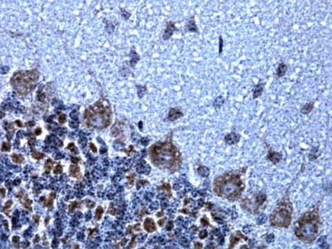
NMDAR1 representative staining pattern/morphology on rat spinal cord at 1:200 dilution. Immunoreactivity is seen as integral membrane and nerve fiber staining of neurons. Frozen section. IHC -Frozen Staining Examples: Rat Spinal Cord

Target description

103-116 kDa

Physical form

Format: Purified
Protein A purified
Purified mouse monoclonal IgG2a liquid in buffer PBS, no preservative.

Storage and Stability

Stable for 1 year at -20ºC from date of receipt.

Analysis Note

Control
Rat brain and spinal cord tissue

Other Notes

Concentration: Please refer to the Certificate of Analysis for the lot-specific concentration.

Legal Information

CHEMICON is a registered trademark of Merck KGaA, Darmstadt, Germany

Disclaimer

Unless otherwise stated in our catalog or other company documentation accompanying the product(s), our products are intended for research use only and are not to be used for any other purpose, which includes but is not limited to, unauthorized commercial uses, in vitro diagnostic uses, ex vivo or in vivo therapeutic uses or any type of consumption or application to humans or animals.

Not finding the right product?  

Try our Product Selector Tool.

Storage Class Code

12 - Non Combustible Liquids

WGK

WGK 2

Flash Point(F)

Not applicable

Flash Point(C)

Not applicable


Certificates of Analysis (COA)

Search for Certificates of Analysis (COA) by entering the products Lot/Batch Number. Lot and Batch Numbers can be found on a product’s label following the words ‘Lot’ or ‘Batch’.

Already Own This Product?

Find documentation for the products that you have recently purchased in the Document Library.

Visit the Document Library

Stroke patients develop antibodies that react with components of N-methyl-D-aspartate receptor subunit 1 in proportion to lesion size.
Kalev-Zylinska, ML; Symes, W; Little, KC; Sun, P; Wen, D; Qiao, L; Young, D; During, MJ; Barber, PA
Stroke null
Csaba Cserép et al.
PloS one, 7(5), e37753-e37753 (2012-06-05)
GABA (gamma-aminobutyric-acid), the main inhibitory neurotransmitter in the adult brain, exerts depolarizing (excitatory) actions during development and this GABAergic depolarization cooperates with NMDARs (N-methyl-D-aspartate receptors) to drive spontaneous synchronous activity (SSA) that is fundamentally important for developing neuronal networks. Although
Catherine L Salussolia et al.
The Journal of neuroscience : the official journal of the Society for Neuroscience, 31(31), 11295-11304 (2011-08-05)
Ionotropic glutamate receptors (iGluRs), including the NMDA receptor subtype, are ligand-gated ion channels critical to fast signaling in the CNS. NMDA receptors are obligate heterotetramers composed of two GluN1 and typically two GluN2 subunits. However, the arrangement of GluN subunits
John E Rash et al.
Journal of neurocytology, 34(3-5), 307-341 (2006-07-15)
Odorant/receptor binding and initial olfactory information processing occurs in olfactory receptor neurons (ORNs) within the olfactory epithelium. Subsequent information coding involves high-frequency spike synchronization of paired mitral/tufted cell dendrites within olfactory bulb (OB) glomeruli via positive feedback between glutamate receptors
Transcriptome analysis of synaptoneurosomes identifies neuroplasticity genes overexpressed in incipient Alzheimer's disease.
Williams, C; Mehrian Shai, R; Wu, Y; Hsu, YH; Sitzer, T; Spann, B; McCleary, C; Mo, Y; Miller, CA
Testing null

Our team of scientists has experience in all areas of research including Life Science, Material Science, Chemical Synthesis, Chromatography, Analytical and many others.

Contact Technical Service