Skip to Content
Merck
All Photos(2)

Key Documents

MAB326R

Sigma-Aldrich

Anti-CNPase Antibody, clone 11-5B

clone 11-5B, Chemicon®, from mouse

Sign Into View Organizational & Contract Pricing


About This Item

UNSPSC Code:
12352203
eCl@ss:
32160702
NACRES:
NA.41
clone:
11-5B, monoclonal
application:
ICC
IHC (p)
WB
species reactivity:
rat, human, bovine, canine, sheep, pig, rabbit, mouse
technique(s):
immunocytochemistry: suitable
immunohistochemistry (formalin-fixed, paraffin-embedded sections): suitable
western blot: suitable
citations:
22

biological source

mouse

Quality Level

antibody form

purified antibody

antibody product type

primary antibodies

clone

11-5B, monoclonal

species reactivity

rat, human, bovine, canine, sheep, pig, rabbit, mouse

manufacturer/tradename

Chemicon®

technique(s)

immunocytochemistry: suitable
immunohistochemistry (formalin-fixed, paraffin-embedded sections): suitable
western blot: suitable

isotype

IgG1

NCBI accession no.

UniProt accession no.

shipped in

wet ice

target post-translational modification

unmodified

Gene Information

human ... CNP(1267)

General description

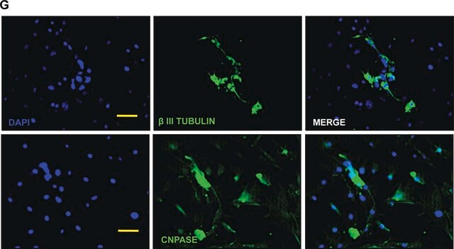
The enzyme 2′, 3′-cyclic nucleotide 3′-phosphodi-esterase (CNP) is expressed at high levels by oligodendrocytes in the central nervous system and by Schwann cells in the peripheral nervous system (Sprinkle, 1989). By virtue of this cell-specific expression, CNP is recognized as a characteristic marker for these two myelin-producing glial cell types (Sprinkle, 1989; Kim et al., 1984; Sheedlo & Sprinkle, 1984; McMorris et al., 1984). Beyond its enzymatic activity of cleaving the 2′, 3′-cyclic terminus of nucleotides (Sprinkle, 1989), the physiological role of CNP is still under investigation. CNP activity has been correlated with myelin and myelin formation, and a dramatic decrease in CNP activity is associated with demyelinating diseases such as multiple sclerosis (Sprinkle, 1989). This enzyme is composed of two proteins, CNP1 (46 kD) and CNP2 (48 kD) (Sprinkle, 1989; Sprinkle et al., 1987). Although the ratio of CNP1/CNP2 may vary from species to species, their shared primary sequence is conserved phyIogenetically. CNP has recently been localized to human chromosome 17 by amplification of somatic cell hybrid DNA using the polymerase chain reaction (PCR) and by Southern blotting of Hind Ill genomic DNA digests (Sprinkle et al., 1991). Since anti-CNP reacts with a highly conserved region of the enzyme, it can be considered as pan-anti-CNP (Sprinkle et al., 1987). Anti-CNP can be used as a marker to identify Schwann cells and oligodendrocytes in cell cuIture and in tissue sections, as well as to localize CNP in cell membrane fractions. As CNP is expressed relatively early in postnatal development, anti-CNP is especially useful for the early identification of oligodendrocytes.

Specificity

Anti-CNP reacts with both CNP1 and CNP2 in many species.

Immunogen

Purified human 2’, 3’-cyclic nucleotide 3’-phosphodiesterase

Application

Immunohistochemistry:
A previous lot of this antibody was used in IH (10 μg/mL antibody, prepared fresh daily).

Immunocytochemistry:
A previous lot of this antibody was used in IC (10 μg/mL antibody, prepared fresh daily).

Optimal working dilutions must be determined by end user.
This Anti-CNPase Antibody, clone 11-5B is validated for use in IC, IH, IH(P), WB for the detection of CNPase.

Quality

Evaluated by Western Blot on Mouse brain lysates.

Western Blotting Analysis:
1:500 dilution of this antibody detected CNPASE 1/2 on 10 µg of Mouse brain lysates.

Target description

48 & 46 kDa

Physical form

Format: Purified
Purified mouse monoclonal IgG1 in buffer containing 0.02M Phosphate buffer, pH 7.6, 0.25M NaCl with 0.1% sodium azide.

Analysis Note

Control
Oligodendrocyte culture, Brain lysate

Other Notes

Concentration: Please refer to the Certificate of Analysis for the lot-specific concentration.

Legal Information

CHEMICON is a registered trademark of Merck KGaA, Darmstadt, Germany

Not finding the right product?  

Try our Product Selector Tool.

Storage Class Code

10 - Combustible liquids

WGK

WGK 2

Flash Point(F)

Not applicable

Flash Point(C)

Not applicable


Certificates of Analysis (COA)

Search for Certificates of Analysis (COA) by entering the products Lot/Batch Number. Lot and Batch Numbers can be found on a product’s label following the words ‘Lot’ or ‘Batch’.

Already Own This Product?

Find documentation for the products that you have recently purchased in the Document Library.

Visit the Document Library

Xiaofei Li et al.
EBioMedicine, 13, 55-65 (2016-11-08)
Stem cells have a high therapeutic potential for the treatment of spinal cord injury (SCI). We have shown previously that endogenous stem cell potential is confined to ependymal cells in the adult spinal cord which could be targeted for non-invasive
Karine Choquet et al.
Molecular brain, 10(1), 13-13 (2017-04-15)
Recessive mutations in the ubiquitously expressed POLR3A gene cause one of the most frequent forms of childhood-onset hypomyelinating leukodystrophy (HLD): POLR3-HLD. POLR3A encodes the largest subunit of RNA Polymerase III (Pol III), which is responsible for the transcription of transfer
Eriola Hoxha et al.
Glia, 69(10), 2419-2428 (2021-06-18)
Elovl5 elongates fatty acids with 18 carbon atoms and in cooperation with other enzymes guarantees the normal levels of very long-chain fatty acids, which are necessary for a proper membrane structure. Action potential conduction along myelinated axons depends on structural
Laurent Methot et al.
Glia, 66(12), 2659-2672 (2018-10-20)
Nuclear factor-kappaB (NF-κB) is activated in neural progenitor cells in the developing murine cerebral cortex during the neurogenic phase, when it acts to prevent premature neuronal differentiation. Here we show that NF-κB activation continues in mouse neocortical neural progenitor cells
Miriam J Schönenberger et al.
Methods in molecular biology (Clifton, N.J.), 1595, 13-26 (2017-04-15)
In the central nervous system (CNS) peroxisomes are present in all cell types, namely neurons, oligodendrocytes, astrocytes, microglia, and endothelial cells. Brain peroxisomes are smaller in size compared to peroxisomes from other tissues and are therefore referred to as microperoxisomes.

Our team of scientists has experience in all areas of research including Life Science, Material Science, Chemical Synthesis, Chromatography, Analytical and many others.

Contact Technical Service