Skip to Content
MilliporeSigma
All Photos(1)

Key Documents

MABF2158

Sigma-Aldrich

Anti-TMPRSS2 Antibody, clone P5H9-A3

clone P5H9-A3, from mouse

Synonym(s):

Transmembrane protease serine 2, Serine protease 10

Sign Into View Organizational & Contract Pricing


About This Item

UNSPSC Code:
12352203
eCl@ss:
32160702
NACRES:
NA.43

biological source

mouse

antibody form

purified antibody

antibody product type

primary antibodies

clone

P5H9-A3, monoclonal

species reactivity

human

packaging

antibody small pack of 25 μg

technique(s)

ELISA: suitable
immunohistochemistry: suitable (paraffin)
western blot: suitable

isotype

IgG1κ

UniProt accession no.

target post-translational modification

unmodified

Gene Information

human ... TMPRSS2(7113)

Related Categories

General description

Transmembrane protease serine 2 (UniProt: O15393; also known as Serine protease 10, TMPRSS2) is encoded by the TMPRSS2 (also known as PRSS10) gene (Gene ID: 7113) in human. TMPRSS2 is a single-pass type II membrane protein of the peptidase S1 family that is shown to proteolytically cleave and activate the viral spike glycoproteins, which facilitate virus-cell membrane fusions. It is shown to facilitate human SARS coronavirus (SARS-CoV) infection via two independent mechanisms, proteolytic cleavage of ACE2 that promotes viral uptake and cleavage of coronavirus spike glycoprotein, which activates the glycoprotein for cathepsin L-independent host cell entry. TMPRSS2 is highly expressed in the prostate tissue and lower expression levels are observed in the epithelia of the colon, stomach, epididymis and breast tissue. Some expression has also been reported in pancreatic acini, hepatic bile ducts, testicular Leydig cells and the kidney. Its expression levels are significantly elevated in both neoplastic prostate and in the epithelium of prostatic hyperplasia. TMPRSS2 has a cytoplasmic domain (aa 1-84), a transmembrane domain (aa 85-105), and an extracellular domain (aa 106-492). Its peptidase S1 domain is localized to amino acids 256-489. It is reported to be proteolytically processed by an autocatalytic mechanism generating the transmembrane protease serine 2 non-catalytic chain (1-255) and the transmembrane protease serine 2 catalytic chain (256-492). Two isoforms of TMPRSS2 have been described that are produced by alternative splicing.

Specificity

Clone P5H9-A3 specifically detects human Transmembrane protease serine 2. It targets an epitope with in the extracellular serine protease domain.

Immunogen

KLH-conjugated linear peptide corresponding to 16 amino acids from the extracellular domain of human transmembrane protease serine 2.

Application

Anti-TMPRSS2, clone P5H9-A3, Cat. No. MABF2158, is a mouse monoclonal antibody that detects Transmembrane protease serine 2 and has been tested for use in ELISA, Immunohistochemistry (Paraffin), and Western Blotting.
Immunohistochemistry (Paraffin) Analysis: 1:50 dilution from a representative lot detected TMPRSS2 in human kidney tissue sections.

Enzyme Immunoassay (ELISA) Analysis: A representative lot detected TMPRSS2 in ELISA applications (Lucas, J.M., et. al. (2008). J Pathol. 215(2):118-25).

Western Blotting Analysis: A representative lot detected TMPRSS2 in Western Blotting applications (Lucas, J.M., et. al. (2008). J Pathol. 215(2):118-25).

Immunohistochemistry (Paraffin) Analysis: A representative lot detected TMPRSS2 in Immunohistochemistry applications (Lucas, J.M., et. al. (2008). J Pathol. 215(2):118-25; Bertram, S., et. al. (2012). PLoS One. 7(4):e35876).
Research Category
Inflammation & Immunology

Quality

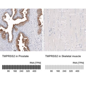
Evaluated by Immunohistochemistry (Paraffin) in human prostate tissue sections.

Immunohistochemistry (Paraffin) Analysis: 1:250 dilution of this antibody detected TMPRSS2 in human prostate tissue sections.

Target description

53.86 kDa calculated.

Physical form

Format: Purified
Protein G purified
Purified mouse monoclonal antibody IgG1 in buffer containing 0.1 M Tris-Glycine (pH 7.4), 150 mM NaCl with 0.05% sodium azide.

Storage and Stability

Stable for 1 year at 2-8°C from date of receipt.

Other Notes

Concentration: Please refer to lot specific datasheet.

Disclaimer

Unless otherwise stated in our catalog or other company documentation accompanying the product(s), our products are intended for research use only and are not to be used for any other purpose, which includes but is not limited to, unauthorized commercial uses, in vitro diagnostic uses, ex vivo or in vivo therapeutic uses or any type of consumption or application to humans or animals.

Not finding the right product?  

Try our Product Selector Tool.


Certificates of Analysis (COA)

Search for Certificates of Analysis (COA) by entering the products Lot/Batch Number. Lot and Batch Numbers can be found on a product’s label following the words ‘Lot’ or ‘Batch’.

Already Own This Product?

Find documentation for the products that you have recently purchased in the Document Library.

Visit the Document Library

Chaitanya Gandikota et al.
Journal of medical virology, 96(4), e29579-e29579 (2024-04-04)
Severe acute respiratory syndrome-related coronavirus 2 (SARS-CoV-2) primarily targets the respiratory system. Physiologically relevant human lung models are indispensable to investigate virus-induced host response and disease pathogenesis. In this study, we generated human induced pluripotent stem cell (iPSC)-derived alveolar organoids (AOs)
Florian Sure et al.
The Journal of biological chemistry, 298(6), 102004-102004 (2022-05-04)
The epithelial sodium channel (ENaC) is a heterotrimer consisting of α-, β-, and γ-subunits. Channel activation requires proteolytic release of inhibitory tracts from the extracellular domains of α-ENaC and γ-ENaC; however, the proteases involved in the removal of the γ-inhibitory
Jessica M Vanslambrouck et al.
Nature communications, 13(1), 5943-5943 (2022-10-09)
While pluripotent stem cell-derived kidney organoids are now being used to model renal disease, the proximal nephron remains immature with limited evidence for key functional solute channels. This may reflect early mispatterning of the nephrogenic mesenchyme and/or insufficient maturation. Here
Laurensius Kevin Lie et al.
Virology, 588, 109889-109889 (2023-10-02)
The lack of suitable in vitro culture model has hampered research on wild-type (WT) human coronaviruses. While 3D tissue or organ cultures have been instrumental for this purpose, such models are challenging, time-consuming, expensive and require extensive cell culture adaptation
Lanlan Zhou et al.
bioRxiv : the preprint server for biology (2020-08-15)
COVID-19 affects vulnerable populations including elderly individuals and patients with cancer. Natural Killer (NK) cells and innate-immune TRAIL suppress transformed and virally-infected cells. ACE2, and TMPRSS2 protease promote SARS-CoV-2 infectivity, while inflammatory cytokines IL-6, or G-CSF worsen COVID-19 severity. We

Our team of scientists has experience in all areas of research including Life Science, Material Science, Chemical Synthesis, Chromatography, Analytical and many others.

Contact Technical Service