Saltar al contenido
MilliporeSigma
Todas las fotos(3)

Key Documents

C2456

Sigma-Aldrich

Monoclonal Anti-Collagen, Type I antibody produced in mouse

clone COL-1, ascites fluid

Sinónimos:

Collagen Type 1 Antibody, Collagen Type 1 Antibody - Monoclonal Anti-Collagen, Type I antibody produced in mouse, Collagen Type I Antibody, Type 1 Collagen Antibody

Iniciar sesiónpara Ver la Fijación de precios por contrato y de la organización


About This Item

MDL number:
UNSPSC Code:
12352203
NACRES:
NA.41

biological source

mouse

Quality Level

conjugate

unconjugated

antibody form

ascites fluid

antibody product type

primary antibodies

clone

COL-1, monoclonal

contains

15 mM sodium azide

species reactivity

bovine, human, pig, rat, rabbit, deer

technique(s)

dot blot: suitable
immunohistochemistry (frozen sections): 1:2000 using human or other mammalian frozen sections
indirect ELISA: suitable

isotype

IgG1

UniProt accession no.

shipped in

dry ice

storage temp.

−20°C

target post-translational modification

unmodified

Gene Information

General description

Collagen type I contributes maximum to the fibrous protein content in mammals. It is also present in arteries and extracellular matrix. The protein has an elongated cylindrical structure with tapering ends. It possess a left-handed helix with three polyproline II-type polypeptide strands.
Type I collagen [1α(I)]2α2 is distributed throughout the body. This fibrillar collagen is found in dermis, bone, tendon, ligament, dentin, fasciae, sclera, cornea, organ capsules and fibrous cartilage. It appears in tissues as the classically designated collagen fibers which are formed from densely-packed thin striated fibrils with marked variation in diameter. Collagen I is synthesized mainly by fibroblasts, osteoblasts, odontoblasts and chondroblasts.Collagen type Iα1 (COL1A1) is encoded by the gene mapped to human chromosome 17q21.33. It is the most abundant extracellular matrix (ECM) protein in humans. Type 1 collagen is the major structural protein of bone, tendon, skin and cornea. The encoded protein is a heterotrimer consisting of two α1-chains and one α2-chain.

Specificity

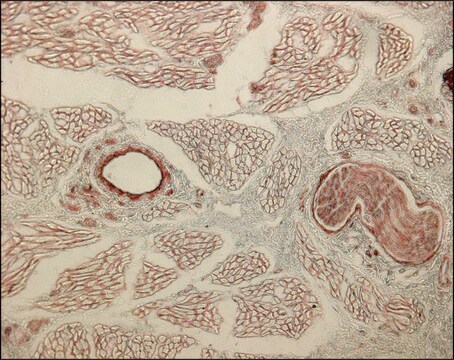
The antibody is reactive with the native (helical) form of collagen type I in ELISA and dot blot. The product is not reactive when tested on thermally denatured molecules. In immunohistochemical staining of acetone-fixed and unfixed frozen sections, a strong staining of connective tissue fibers is seen. Using the dot blot and ELISA techniques, the antibody shows no cross-reactivity with collagen types, II, III, IV, V, VI, VII, IX, X and XI. The epitope recognized by the antibody is sensitive to routine formalin fixation and paraffin embedding.

Immunogen

Bovine skin collagen type I

Application

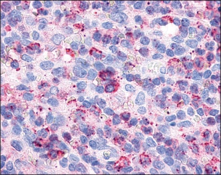
Monoclonal Anti-Collagen Type I may be used for the localization of type I collagen using various immunochemical assays including ELISA, dot blot and immunohistochemistry. A minimum working dilution of 1:2,000 is determined by indirect immunofluorescent staining of human or other mammalian frozen sections.
Monoclonal Anti-Collagen, Type I antibody produced in mouse has been used in:
  • immunohistochemistry
  • dot blot technique
  • indirect immunofluorescence staining

Biochem/physiol Actions

Collagen type I is responsible for mechanical stability, elasticity, strength and toughness, observed in tendons, ligaments, skin, cornea, bone, dentin and many other tissues.
Type 1 collagen serves a structural role in the extracellular matrix by providing mechanical support and resistance to tension. Some of the more important genetic diseases, directly or indirectly involving this collagen type include the majority cases of osteogenesis imperfecta and certain types of Ehlers Danlos syndrome. The development of antibodies against collagens has provided a powerful method for examining the distribution of these connective tissue proteins and for investigation of epithelial-mesenchymal interactions, tumorigenesis and basement membrane biology in ontogeny and epithelial differentiation.

Physical form

Liquid

Storage and Stability

Store at 2-8 °C for up to one month. For extended storage freeze in working aliquots. Repeated freezing and thawing is not recommended. Storage in "frost-free" freezers is not recommended. If slight turbidity occurs upon prolonged storage, clarify the solution by centrifugation before use.

Disclaimer

Unless otherwise stated in our catalog or other company documentation accompanying the product(s), our products are intended for research use only and are not to be used for any other purpose, which includes but is not limited to, unauthorized commercial uses, in vitro diagnostic uses, ex vivo or in vivo therapeutic uses or any type of consumption or application to humans or animals.

¿No encuentra el producto adecuado?  

Pruebe nuestro Herramienta de selección de productos.

Storage Class

12 - Non Combustible Liquids

wgk_germany

nwg

flash_point_f

Not applicable

flash_point_c

Not applicable


Certificados de análisis (COA)

Busque Certificados de análisis (COA) introduciendo el número de lote del producto. Los números de lote se encuentran en la etiqueta del producto después de las palabras «Lot» o «Batch»

¿Ya tiene este producto?

Encuentre la documentación para los productos que ha comprado recientemente en la Biblioteca de documentos.

Visite la Librería de documentos

Gene expressions of small leucine-rich repeat proteoglycans and fibulin-5 are decreased in pelvic organ prolapse
Soderberg M W, et al.
Molecular Human Reproduction, 15(4), 251-257 (2009)
Gemma Pascual et al.
PloS one, 8(11), e80647-e80647 (2013-11-16)
Composite biomaterials designed for the repair of abdominal wall defects are composed of a mesh component and a laminar barrier in contact with the visceral peritoneum. This study assesses the behaviour of a new composite mesh by comparing it with
Collagen fibrillar structure and hierarchies
Collagen: Structure and Mechanics, 49-80 (2008)
Collagen: structure and mechanics, an introduction
Fratzl P.
Collagen: Structure and Mechanics, 1-13 (2008)
Pan Yeung et al.
PloS one, 10(12), e0144139-e0144139 (2015-12-15)
There is a growing trend for researchers to use in vitro 3D models in cancer studies, as they can better recapitulate the complex in vivo situation. And the fact that the progression and development of tumor are closely associated to

Nuestro equipo de científicos tiene experiencia en todas las áreas de investigación: Ciencias de la vida, Ciencia de los materiales, Síntesis química, Cromatografía, Analítica y muchas otras.

Póngase en contacto con el Servicio técnico