Skip to Content
MilliporeSigma
All Photos(1)

Key Documents

06-596

Sigma-Aldrich

Anti-STAT3 Antibody

Upstate®, from rabbit

Synonym(s):

Acute-phase response factor, DNA-binding protein APRF, signal transducer and activator of transcription, signal transducer and activator of transcription, (acute-phase response factor)

Sign Into View Organizational & Contract Pricing


About This Item

UNSPSC Code:
12352203
eCl@ss:
32160702
NACRES:
NA.41

biological source

rabbit

Quality Level

antibody form

purified immunoglobulin

antibody product type

primary antibodies

clone

polyclonal

species reactivity

mouse, human, rat

manufacturer/tradename

Upstate®

technique(s)

ChIP: suitable
electrophoretic mobility shift assay: suitable
immunocytochemistry: suitable
immunoprecipitation (IP): suitable
western blot: suitable

isotype

IgG

NCBI accession no.

UniProt accession no.

shipped in

wet ice

target post-translational modification

unmodified

Gene Information

human ... STAT3(6774)
mouse ... Stat3(20848)

General description

STAT proteins (Signal Transduction and Activators of Transcription) are latent cytoplasmic transcription factors that have the dual function of signal transduction and activation of transcription. STATs are activated by tyrosine phosphorylation in response to different ligands, after which they translocate to the cell nucleus. The N-terminal region is highly homologous among the STAT proteins and surrounds a completely conserved arginine residue. STATs are a part of the JAK-STAT signaling pathway – a major pathway of the immune system. All cytokines transduce critical signals through this pathway. STAT3 has been shown to be activated by IFN alpha but not IFN beta. The transcription factors associated with STAT3 are cJun and cyclic AMP responsive enhancer binding protein (CREB). Deletion of the STAT3 gene in knock out mice was lethal at the early embryonic stage.

Specificity

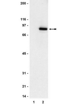
Recognizes STAT3, Mr 92 kDa. Additional unknown bands may be detected.

Immunogen

Bacterially expressed GST fusion protein corresponding to a.a. 688-722 of human STAT 3 (RPESQEHPEADPGSAAPYLKTKFICVTPTTCSNTI). The immunizing sequence is identical in mouse and rat STAT3. The first 28 amino acids are identical to mouse STAT3B.
Epitope: a.a. 688-722

Application

Anti-STAT3 Antibody is a Rabbit Polyclonal Antibody for detection of STAT3 also known as Acute-phase response factor or DNA-binding protein APRF & has been validated in ChIP, EMSA, ICC, IP & WB.
Immunoprecipitation:
4 μg of a previous lot immunoprecipitated STAT3 from 500 μg of EGF-stimulated A431 RIPA lysate.
Gel Shift Assay:
An independent laboratory has reported that this antibody supershifts.
Immunocytochemistry:
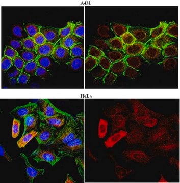
10 μg/mL of a previous lot of this antibody showed positive immunostaining for STAT3 in A431 cells fixed with 95% ethanol, 5% acetic acid.
Research Category
Epigenetics & Nuclear Function
Research Sub Category
Transcription Factors

Quality

Routinely evaluated by western blot on RIPA lysates from EGF stimulated human A431 cells, mouse WEHI or rat L6.

Western Blot Analysis:
0.5-2 μg/mL of this lot detected STAT3 in RIPA lysates from EGF stimulated human A431 cells and previously from mouse WEHI and rat L6.

Target description

92 kDa

Linkage

Replaces: 04-1014

Physical form

Format: Purified
Protein A purified
Protein A purified rabbit IgG in 200 L of 0.1 M Tris-glycine, pH 7.4, 0.15 M NaCl, 0.05% sodium azide. Frozen at -20°C.

Storage and Stability

Stable for 1 year at -20°C from date of receipt.
Handling Recommendations: Upon first thaw, and prior to removing the cap, centrifuge the vial and gently mix the solution. Aliquot into microcentrifuge tubes and store at -20°C. Avoid repeated freeze/thaw cycles, which may damage IgG and affect product performance.

Analysis Note

Control
Positive Antigen Control: Catalog #12-302, EGF-stimulated A431 cell lysate. Add 2.5µL of 2-mercaptoethanol/100µL of lysate and boil for 5 minutes to reduce the preparation. Load 20µg of reduced lysate per lane for minigels.

Other Notes

Concentration: Please refer to the Certificate of Analysis for the lot-specific concentration.

Legal Information

UPSTATE is a registered trademark of Merck KGaA, Darmstadt, Germany

Disclaimer

Unless otherwise stated in our catalog or other company documentation accompanying the product(s), our products are intended for research use only and are not to be used for any other purpose, which includes but is not limited to, unauthorized commercial uses, in vitro diagnostic uses, ex vivo or in vivo therapeutic uses or any type of consumption or application to humans or animals.

Not finding the right product?  

Try our Product Selector Tool.

Storage Class

12 - Non Combustible Liquids

wgk_germany

WGK 1

flash_point_f

Not applicable

flash_point_c

Not applicable


Certificates of Analysis (COA)

Search for Certificates of Analysis (COA) by entering the products Lot/Batch Number. Lot and Batch Numbers can be found on a product’s label following the words ‘Lot’ or ‘Batch’.

Already Own This Product?

Find documentation for the products that you have recently purchased in the Document Library.

Visit the Document Library

Differentiation of oligodendroglial progenitors derived from cortical multipotent cells requires extrinsic signals including activation of gp130/LIFbeta receptors
Marmur, R., et al
The Journal of Neuroscience, 18, 9800-9811 (1998)
M Ernst et al.
The Journal of biological chemistry, 274(14), 9729-9737 (1999-03-27)
Cell type-specific responses to the leukemia inhibitory factor (LIF)/interleukin 6 cytokine family are mediated by dimerization of the LIF receptor alpha-chain (LIFRalpha) with the signal transducer gp130 or of two gp130 molecules followed by activation of the JAK/STAT and Ras/mitogen-activated
The box-1 region of the leukemia inhibitory factor receptor alpha-chain cytoplasmic domain is sufficient for hemopoietic cell proliferation and differentiation
Zhang, Y., et al
The Journal of Biological Chemistry, 273, 34370-34383 (1998)
Kirsten P Stone et al.
Cellular physiology and biochemistry : international journal of experimental cellular physiology, biochemistry, and pharmacology, 28(1), 115-124 (2011-08-26)
Interleukin (IL)-15 and its receptors are induced by tumor necrosis factor α (TNF) in the cerebral endothelial cells composing the blood-brain barrier, but it is not yet clear how IL-15 modulates endothelial function. Contrary to the known induction of JAK/STAT3
Nuclear expression of a group II intron is consistent with spliceosomal intron ancestry.
Chalamcharla VR, Curcio MJ, Belfort M
Genes & Development null

Our team of scientists has experience in all areas of research including Life Science, Material Science, Chemical Synthesis, Chromatography, Analytical and many others.

Contact Technical Service