Skip to Content
Merck
All Photos(4)

Documents

S5768

Sigma-Aldrich

Monoclonal Anti-Synaptophysin antibody produced in mouse

clone SVP-38, ascites fluid

Synonym(s):

Synaptophysin Antibody, Synaptophysin Antibody - Monoclonal Anti-Synaptophysin antibody produced in mouse

Sign Into View Organizational & Contract Pricing


About This Item

MDL number:
UNSPSC Code:
12352203
NACRES:
NA.41

biological source

mouse

Quality Level

conjugate

unconjugated

antibody form

ascites fluid

antibody product type

primary antibodies

clone

SVP-38, monoclonal

mol wt

antigen 38 kDa

contains

15 mM sodium azide

species reactivity

pig, rat, human, guinea pig

technique(s)

immunohistochemistry (formalin-fixed, paraffin-embedded sections): suitable
immunohistochemistry (frozen sections): 1:200 using rat cerebellum
indirect ELISA: suitable
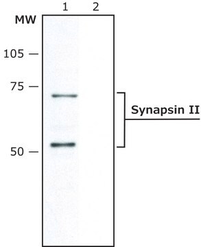
western blot: suitable

isotype

IgG1

UniProt accession no.

shipped in

dry ice

storage temp.

−20°C

target post-translational modification

unmodified

Gene Information

human ... SYP(6855)
rat ... Syp(24804)

General description

Synaptophysin (SYP) is an integral membrane glycoprotein of 38,000 Da and it has four membrane-spanning domains. Synaptophysin is localized to neurons, neuromuscular junctions, paraganglia cells, hypophysis, pancreatic islet cells, and adrenal cells. SYP gene is mapped to human chromosome Xp11.23. Monoclonal Anti-Synaptophysin (mouse IgG1 isotype) is derived from the hybridoma produced by the fusion of mouse myeloma cells and splenocytes from an immunized mouse.

Specificity

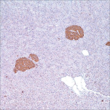
Labels synaptophysin in neurons, neuromuscular junctions, benign and malignant neural tumors, paraganglia cells, hypophysis, pancreatic islet cells, and adrenal cells.

Immunogen

Rat retina synaptosome.

Application

Monoclonal Anti-Synaptophysin antibody has been used in western blotting.

Biochem/physiol Actions

Synaptophysin participates in calcium dependent exocytosis. The antibody detects synaptophysin in many types of benign and malignant neural and epithelial neuroendocrine neoplasms. It is widely used as a potential marker to study the circulation of synapses in the brain and to discover the basic features of the life cycle of synaptic vesicles (SVs).

Physical form

Monoclonal Anti-Synaptophysin is provided as ascites fluid containing 0.1% sodium azide.

Storage and Stability

For continuous use, store at 2-8 °C for up to one month. For extended storage, the solution may be frozen in working aliquots. Repeated freezing and thawing is not recommended. Storage in "frost-free" freezers is not recommended. If slight turbidity occurs upon prolonged storage, clarify by centrifugation before use.

Disclaimer

Unless otherwise stated in our catalog or other company documentation accompanying the product(s), our products are intended for research use only and are not to be used for any other purpose, which includes but is not limited to, unauthorized commercial uses, in vitro diagnostic uses, ex vivo or in vivo therapeutic uses or any type of consumption or application to humans or animals.

Not finding the right product?  

Try our Product Selector Tool.

Storage Class Code

12 - Non Combustible Liquids

WGK

WGK 2

Flash Point(F)

Not applicable

Flash Point(C)

Not applicable


Certificates of Analysis (COA)

Search for Certificates of Analysis (COA) by entering the products Lot/Batch Number. Lot and Batch Numbers can be found on a product’s label following the words ‘Lot’ or ‘Batch’.

Already Own This Product?

Find documentation for the products that you have recently purchased in the Document Library.

Visit the Document Library

Synaptophysin: leading actor or walk-on role in synaptic vesicle exocytosis?
Valtorta F
Bioessays, 26(4), 445-453 (2004)
Coordinate action of pre-and postsynaptic brain-derived neurotrophic factor is required for AMPAR trafficking and acquisition of in vitro classical conditioning.
Li W and Keifer J
Neuroscience, 155(3), 686-697 (2008)
Synaptophysin: leading actor or walk-on role in synaptic vesicle exocytosis?
Valtorta F, et al.
Bioessays, 26(4), 445-453 (2004)
Synaptophysin: a marker protein for neuroendocrine cells and neoplasms.
Wiedenmann B, et al.
Proceedings of the National Academy of Sciences of the USA, 83(10), 3500-3504 (1986)
The distribution of synapsin I and synaptophysin in hippocampal neurons developing in culture.
Fletcher TL, et al.
The Journal of Neuroscience, 11(6), 1617-1626 (1991)

Our team of scientists has experience in all areas of research including Life Science, Material Science, Chemical Synthesis, Chromatography, Analytical and many others.

Contact Technical Service