Skip to Content
Merck
All Photos(1)

Documents

M8177

Sigma-Aldrich

Anti-p38 MAP Kinase, Activated (Diphosphorylated p38) antibody, Mouse monoclonal

clone P38-TY, purified from hybridoma cell culture, buffered aqueous solution

Synonym(s):

Anti-MAP Kinase, p38, Activated, Monoclonal Anti-p38 MAP Kinase, Activated (Diphosphorylated p38) antibody produced in mouse

Sign Into View Organizational & Contract Pricing


About This Item

MDL number:
UNSPSC Code:
12352203
NACRES:
NA.44

biological source

mouse

Quality Level

conjugate

unconjugated

antibody form

purified from hybridoma cell culture

antibody product type

primary antibodies

clone

P38-TY, monoclonal

form

buffered aqueous solution

species reactivity

rat, human, mouse

concentration

~2 mg/mL

technique(s)

immunocytochemistry: suitable
indirect ELISA: suitable
microarray: suitable
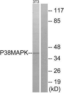
western blot: 1 μg/mL using whole cell extract from a rat fibroblast cell line, Rat1, activated with sorbitol

isotype

IgG2a

UniProt accession no.

shipped in

dry ice

storage temp.

−20°C

target post-translational modification

unmodified

Gene Information

human ... MAPK14(1432)
mouse ... Mapk14(26416)
rat ... Mapk14(81649)

General description

Monoclonal Anti-p38 MAP Kinase, Activated (Diphosphorylated p38) (mouse IgG2a isotype) is derived from the P38-TY hybridoma produced by the fusion of mouse myeloma cells and splenocytes from BALB/c mice. The p38 mitogen-activated protein kinases (MAPK) comprises the stress-activated protein kinase (SAPK) 2b, SAPK3 and SAPK4 (also termed p38β-δ). All these kinases contain a glycine residue in their TXY(Thr−Xaa−Tyr motif) activation motif.

Specificity

Reacts specifically with the active diphosphorylated form of p38 MAP kinase and its closely related isoforms. Does not recognize the non-phosphorylated and mono­phosphory­lated forms of the p38 MAP kinase molecule, or the non-phosphorylated, monophosphorylated and diphosphorylated forms of JNK and ERK-MAP kinases. The epitope recognized by the antibody contains the phosphorylated threonine and tyrosine residues within the regulatory site of active p38 MAP kinase (Thr180-Gly181-Tyr182).

Immunogen

Synthetic peptide HTDDEMpTGpYVATR corresponding to the phosphorylated form of p38 MAP kinase activation loop, coupled to KLH.

Application

Monoclonal Anti-p38 MAP Kinase, Activated (Diphosphorylated p38) antibody produced in mouse may be used in immunoblotting, enzyme linked immunosorbent assay (ELISA), immunostaining, western blotting, immunohistochemistry and in immunocytochemistry.

Biochem/physiol Actions

The p38 mitogen-activated protein kinases (MAPK) are activated by phosphorylation of both tyrosine (Y) and threonine (T) residues organized in a TXY motif. The residue in between the two phosphorylated residues determines the specificity of activation of the MAPKs, and is glutamic acid for extracellular-signal regulated kinase (ERK) (TEY), proline for c-Jun N-terminal kinases (JNK) and glycine for p38 MAPK. Phosphorylation of both tyrosine and threonine is essential for the full activation of all MAPKs. ERK generally transmits signals leading to cell proliferation, p38 MAPK and JNK are both mostly stress-responsive kinases3 and have been implicated in cell death in several cellular systems. Antibodies reacting specifically with activated p38 MAPK are useful tools in the study of the intracellular location of p38 MAPK enzymes, and in sorting out the signal transduction pathways of the MAPK superfamily.

Physical form

Solution in 0.01 M phosphate buffered saline, pH 7.4, containing 15 mM sodium azide.

Preparation Note

From a culture supernatant of bioreactor grown hybridoma.

Disclaimer

Unless otherwise stated in our catalog or other company documentation accompanying the product(s), our products are intended for research use only and are not to be used for any other purpose, which includes but is not limited to, unauthorized commercial uses, in vitro diagnostic uses, ex vivo or in vivo therapeutic uses or any type of consumption or application to humans or animals.

Not finding the right product?  

Try our Product Selector Tool.

Storage Class Code

10 - Combustible liquids

WGK

nwg

Flash Point(F)

Not applicable

Flash Point(C)

Not applicable


Certificates of Analysis (COA)

Search for Certificates of Analysis (COA) by entering the products Lot/Batch Number. Lot and Batch Numbers can be found on a product’s label following the words ‘Lot’ or ‘Batch’.

Already Own This Product?

Find documentation for the products that you have recently purchased in the Document Library.

Visit the Document Library

Lucinda Rand et al.
Journal of immunology (Baltimore, Md. : 1950), 182(9), 5865-5872 (2009-04-22)
Mycobacterium tuberculosis (M. tb) must cause lung disease to spread. Matrix metalloproteinases (MMPs) degrade the extracellular matrix and are implicated in tuberculosis-driven tissue destruction. We investigated signaling pathways regulating macrophage MMP-1 and -7 in human pulmonary tuberculosis and examine the
Simultaneous measurement of ERK, p38, and JNK MAP kinase cascades in vascular smooth muscle cells
Chevalier D, et al.
Journal of Pharmacological and Toxicological Methods, 44(2), 429-439 (2000)
Gian Felice DeNicola et al.
Nature structural & molecular biology, 20(10), 1182-1190 (2013-09-17)
p38α mitogen-activated protein kinase (p38α) is activated by a variety of mechanisms, including autophosphorylation initiated by TGFβ-activated kinase 1 binding protein 1 (TAB1) during myocardial ischemia and other stresses. Chemical-genetic approaches and coexpression in mammalian, bacterial and cell-free systems revealed
Matrix metalloproteinase-1 is regulated in tuberculosis by a p38 MAPK-dependent, p-aminosalicylic acid-sensitive signaling cascade
Rand L, et al.
Journal of Immunology, 182(9), 5865-5872 (2009)
Gian F De Nicola et al.
JCI insight, 3(16) (2018-08-24)
Inhibiting MAPK14 (p38α) diminishes cardiac damage in myocardial ischemia. During myocardial ischemia, p38α interacts with TAB1, a scaffold protein, which promotes p38α autoactivation; active p38α (pp38α) then transphosphorylates TAB1. Previously, we solved the X-ray structure of the p38α-TAB1 (residues 384-412)

Our team of scientists has experience in all areas of research including Life Science, Material Science, Chemical Synthesis, Chromatography, Analytical and many others.

Contact Technical Service