Skip to Content
Merck
All Photos(1)

Key Documents

17-344

Sigma-Aldrich

H2A.X Phosphorylation Assay Kit (Flow Cytometry)

The H2A.X Phosphorylation Assay Kit (Flow cytometry) is a cell based assay formatted for flow cytometric detection of levels of phosphorylated Histone H2A.X.

Synonym(s):

Assay for H2A.X, Flow Cytometry Assay Kit, H2A.X Phosphorylation Kit

Sign Into View Organizational & Contract Pricing


About This Item

UNSPSC Code:
12161503
eCl@ss:
32161000
NACRES:
NA.32

Quality Level

manufacturer/tradename

Upstate®

technique(s)

activity assay: suitable
flow cytometry: suitable

NCBI accession no.

UniProt accession no.

detection method

fluorometric

shipped in

dry ice

Related Categories

General description

Phosphorylation of the histone variant H2A.X is a rapid and sensitive response to double strand DNA breaks. The H2A.X Phosphorylation Assay Kit (Flow cytometry) is a cell based assay formatted for flow cytometric detection of levels of phosphorylated histone H2A.X.

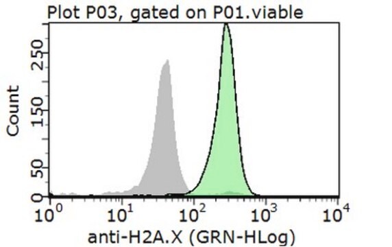
The H2A.X Phosphorylation Assay Kit (Flow cytometry) is a cell based assay formatted for flow cytometric detection of levels of phosphorylated Histone H2A.X. Cells are cultured in microplates, treated with agents that induce DNA damage or apoptosis, which stimulates H2A.X phosphorylation. Cells are then fixed and permeabilized in preparation for staining and detection. Histone H2A.X phosphorylated at serine 139 is detected by the addition of the anti-phospho-Histone H2A.X, FITC conjugate. Cells are then scanned in a flow cytometer to quantitate the number of cells staining positive for phosphorylated Histone H2A.X.

Application

Research Category
Neuroscience
Research Sub Category
Neurodegenerative Diseases

Packaging

100 assays

Components

Anti-phospho H2A.X, FITC conjugate

Normal Mouse IgG, FITC conjugate (Cat.# 12-487)

16X Fixation Solution

10X Permeabilization Solution

10X Wash Solution

Quality

phosphorylation of Histone H2A.X

Legal Information

UPSTATE is a registered trademark of Merck KGaA, Darmstadt, Germany

Disclaimer

Unless otherwise stated in our catalog or other company documentation accompanying the product(s), our products are intended for research use only and are not to be used for any other purpose, which includes but is not limited to, unauthorized commercial uses, in vitro diagnostic uses, ex vivo or in vivo therapeutic uses or any type of consumption or application to humans or animals.

Signal Word

Danger

Hazard Classifications

Acute Tox. 2 Inhalation - Acute Tox. 3 Dermal - Acute Tox. 3 Oral - Carc. 1B - Eye Dam. 1 - Flam. Liq. 2 - Muta. 2 - Skin Corr. 1B - Skin Sens. 1 - STOT SE 1 - STOT SE 3

Target Organs

Eyes,Central nervous system, Respiratory system

Storage Class Code

3 - Flammable liquids


Certificates of Analysis (COA)

Search for Certificates of Analysis (COA) by entering the products Lot/Batch Number. Lot and Batch Numbers can be found on a product’s label following the words ‘Lot’ or ‘Batch’.

Already Own This Product?

Find documentation for the products that you have recently purchased in the Document Library.

Visit the Document Library

Yu Wang et al.
Mutation research, 734(1-2), 20-29 (2012-05-09)
Berberine has been shown to possess anti-tumor activity against a wide spectrum of cancer cells. It inhibits cancer cell proliferation by inducing cell cycle arrest, at G1 and/or G2/M, and apoptosis. While it has been documented that berberine induces G1
Runtao Zhong et al.
RSC advances, 10(49), 29311-29319 (2020-08-07)
Immunofluorescence (IF) is a common method used in cell biology. The conventional protocol for IF staining is time and labor-intensive, operator dependent and reagent-consuming. Magnetic Bead (MB)-based microdevices are frequently utilized in cellular assays, but integration of simple and efficient
Angelo Fortunato et al.
PLoS biology, 19(11), e3001471-e3001471 (2021-11-18)
Trichoplax adhaerens is the simplest multicellular animal with tissue differentiation and somatic cell turnover. Like all other multicellular organisms, it should be vulnerable to cancer, yet there have been no reports of cancer in T. adhaerens or any other placozoan.
Amy D Guertin et al.
Molecular cancer therapeutics, 12(8), 1442-1452 (2013-05-24)
Inhibition of the DNA damage checkpoint kinase WEE1 potentiates genotoxic chemotherapies by abrogating cell-cycle arrest and proper DNA repair. However, WEE1 is also essential for unperturbed cell division in the absence of extrinsic insult. Here, we investigate the anticancer potential
Amy D Guertin et al.
Cancer cell international, 12(1), 45-45 (2012-11-15)
Inhibition of kinases involved in the DNA damage response sensitizes cells to genotoxic agents by abrogating checkpoint-induced cell cycle arrest. CHK1 and WEE1 act in a pathway upstream of CDK1 to inhibit cell cycle progression in response to damaged DNA.

Our team of scientists has experience in all areas of research including Life Science, Material Science, Chemical Synthesis, Chromatography, Analytical and many others.

Contact Technical Service