Skip to Content
Merck
All Photos(2)

Key Documents

SAB4200763

Sigma-Aldrich

Anti-Dystrophin antibody, Mouse monoclonal

enhanced validation

clone MANDRA1, purified from hybridoma cell culture

Synonym(s):

Anti-DMD

Sign Into View Organizational & Contract Pricing


About This Item

UNSPSC Code:
12352203
NACRES:
NA.41

biological source

mouse

Quality Level

antibody form

purified from hybridoma cell culture

antibody product type

primary antibodies

clone

MANDRA1, monoclonal

form

buffered aqueous solution

mol wt

~427 kDa

species reactivity

zebrafish, rat, mouse, human

enhanced validation

independent
Learn more about Antibody Enhanced Validation

concentration

~1.0 mg/mL

technique(s)

immunoblotting: suitable
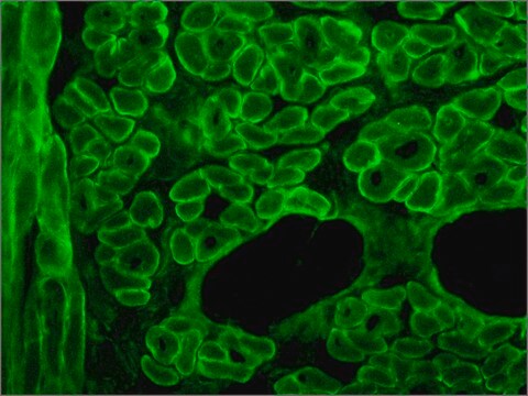
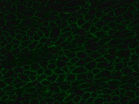
immunofluorescence: suitable
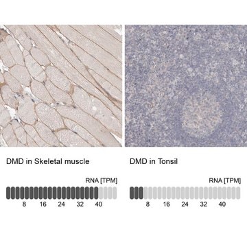
immunohistochemistry: 10-20 μg/mL using acetone fixed rat tongue frozen sections

isotype

IgG1

UniProt accession no.

shipped in

dry ice

storage temp.

−20°C

target post-translational modification

unmodified

Gene Information

human ... DMD(1756)

General description

Anti-Dystrophin antibody, Mouse monoclonal (mouse IgG1 isotype) is derived from the MANDRA1 hybridoma produced by the fusion of mouse myeloma cells and splenocytes from BALB/c mice. Dystrophin is a rod-shaped cytoskeletal protein located to the periphery (plasma membrane) of normal striated muscle fibers.
Dystrophin is encoded by the gene mapped to human chromosome Xp21.2-p21.1.

Specificity

Anti-Dystrophin antibody, Mouse monoclonal specifically recognizes an epitope located on the 128 amino acids at the end of the C-terminal domain of the human Dystrophin molecule (residues 3558-3684). The antibody reacts with Dystrophin from human, mouse, rat and zebrafish origin. The antibody is specific to dystrophin and does not react with -actinin or utrophin.

Application

Anti-Dystrophin antibody, Mouse monoclonal has been used in:
  • immunohistochemistry
  • immunoblotting
  • immunofluorescence
  • enzyme-linked immunosorbent assay (ELISA)

Biochem/physiol Actions

Dystrophin is absent, reduced or altered because of mutation in Duchenne and Becker muscular dystrophies (DMD/BMD) or in its homologue in mouse. Severe DMD is associated with a marked Dystrophin deficiency whereas patients with the milder form of BMD show less pronounced abnormalities of protein expression.

Physical form

Solution in 0.01 M phosphate buffered saline, pH 7.4, containing 15 mM sodium azide.

Storage and Stability

For continuous use, store at 2–8 °C for up to one month. For extended storage, freeze in working aliquots. Repeated freezing and thawing is not recommended. If slight turbidity occurs upon prolonged storage, clarify the solution by centrifugation before use. Working dilution samples should be discarded if not used within 12 hours.

Disclaimer

Unless otherwise stated in our catalog, our products are intended for research use only and are not to be used for any other purpose, which includes but is not limited to, unauthorized commercial uses, in vitro diagnostic uses, ex vivo or in vivo therapeutic uses or any type of consumption or application to humans or animals.

Not finding the right product?  

Try our Product Selector Tool.

Storage Class Code

10 - Combustible liquids

Flash Point(F)

Not applicable

Flash Point(C)

Not applicable


Certificates of Analysis (COA)

Search for Certificates of Analysis (COA) by entering the products Lot/Batch Number. Lot and Batch Numbers can be found on a product’s label following the words ‘Lot’ or ‘Batch’.

Already Own This Product?

Find documentation for the products that you have recently purchased in the Document Library.

Visit the Document Library

Duchenne muscular dystrophy and dystrophin: pathogenesis and opportunities for treatment
Nowak KJ and Davies KE
EMBO Reports, 5(9), 872-876 (2004)
Analysis of dystrophin gene deletions by multiplex PCR in Moroccan patients
Sbiti A, et al.
BioMed Research International, 2(3), 158-160 (2002)
Davide Rovina et al.
Stem cell research, 40, 101544-101544 (2019-08-30)
Duchenne's muscular dystrophy (DMD) is a neuromuscular disorder affecting skeletal and cardiac muscle function, caused by mutations in the dystrophin (DMD) gene. Dermal fibroblasts, isolated from a DMD patient with a reported deletion of exons 51 to 53 in the

Our team of scientists has experience in all areas of research including Life Science, Material Science, Chemical Synthesis, Chromatography, Analytical and many others.

Contact Technical Service