Skip to Content
Merck
All Photos(4)

Documents

MAB1568

Sigma-Aldrich

Anti-Calretinin Antibody, clone 6B8.2

clone 6B8.2, Chemicon®, from mouse

Synonym(s):

calbindin 2, calbindin 2, (29kD, calretinin)

Sign Into View Organizational & Contract Pricing


About This Item

UNSPSC Code:
12352203
eCl@ss:
32160702
NACRES:
NA.41

biological source

mouse

Quality Level

antibody form

purified immunoglobulin

antibody product type

primary antibodies

clone

6B8.2, monoclonal

species reactivity

mouse, rat

manufacturer/tradename

Chemicon®

technique(s)

immunohistochemistry: suitable
western blot: suitable

isotype

IgG1

NCBI accession no.

UniProt accession no.

shipped in

wet ice

target post-translational modification

unmodified

Gene Information

human ... CALB2(794)

General description

Calretinin is a 29 kDa calcium binding protein that is expressed in central and peripheral nervous system and in many normal and pathological tissues. Calretinin is also considered an important diagnostic aid in the differential diagnosis of cystic and solid ameloblastic tumors. The rat and human calretinin exhibit 98% sequence homology and 91% homology to many other species. Calretinin is generally expressed with two other calcium binding proteins Calbindin D-29 and parvalbumin in CNS and other peripheral tissues. Calretinin antibodies have been used to detect a number of carcinomas by clinicopathological, immunohistochemical techniques including, epithelial malignant mesotheliomas, Ovarian yolk sac tumor, Adenomatoid tumors of the uterus etc

Specificity

Calretinin is a neuron specific calcium binding protein (31 kDa) found mostly in the CNS and retina. It is present in subsets of neurons throughout the brain and spinal cord, including sensory ganglia. It shares about 55% amino acid homology with calbindin, another calcium binding protein found in brain and periphery. Studies suggest that calretinin, like calbindin, may be neuroprotective.
Other species have not been tested.

Immunogen

Recombinant rat calretinin (Heinze, L., 2007).

Application

Anti-Calretinin Antibody, clone 6B8.2 detects level of Calretinin & has been published & validated for use in IH & WB.
Immunohistochemistry:
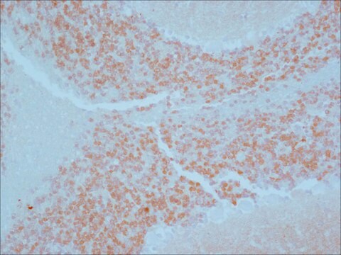
On rat brain tissue:
IFA: 1:1,000-1:5,000
PAP: 1:1,000-1:5,000
The antibody has been used successfully on formalin-fixed, paraffin embedded human tissue after pre-treatment of the tissue slides with citrate buffer and heat treatment. Suggested starting dilution 1:100-1:400. Proteolytic digestion with pepsin is detrimental to the immunostaining.

Western blot:
1:1,000-1:3,000

Optimal working dilutions must be determined by the end user.
Research Category
Neuroscience
Research Sub Category
Signaling Neuroscience

Quality

Routinely evaluated by Western Blot on mouse brain lysates.

Western Blot Analysis:
1:1000 dilution of this lot detected Calretinin on 10 μg of mouse brain lysates.

Target description

31 kDa

Physical form

Format: Purified
Protein A purified
Purified mouse monoclonal IgG1 in buffer containing 0.02 M Phosphate buffer, 0.25 M NaCl, pH 7.6 with 0.1% sodium azide.

Storage and Stability

Stable for 6 months at 2-8°C in undiluted aliquots from date of receipt.

Analysis Note

Control
Rat brain cortex, Mesothelioma tissue, mouse brain lysate.

Other Notes

Concentration: Please refer to the Certificate of Analysis for the lot-specific concentration.

Legal Information

CHEMICON is a registered trademark of Merck KGaA, Darmstadt, Germany

Disclaimer

Unless otherwise stated in our catalog or other company documentation accompanying the product(s), our products are intended for research use only and are not to be used for any other purpose, which includes but is not limited to, unauthorized commercial uses, in vitro diagnostic uses, ex vivo or in vivo therapeutic uses or any type of consumption or application to humans or animals.

Not finding the right product?  

Try our Product Selector Tool.

Storage Class Code

10 - Combustible liquids

WGK

WGK 2

Flash Point(F)

Not applicable

Flash Point(C)

Not applicable


Certificates of Analysis (COA)

Search for Certificates of Analysis (COA) by entering the products Lot/Batch Number. Lot and Batch Numbers can be found on a product’s label following the words ‘Lot’ or ‘Batch’.

Already Own This Product?

Find documentation for the products that you have recently purchased in the Document Library.

Visit the Document Library

Igor Jakovcevski et al.
Cerebral cortex (New York, N.Y. : 1991), 21(8), 1771-1782 (2010-12-09)
Cortical γ-aminobutyric acid (GABA)ergic interneurons in rodents originate mainly in ventrally positioned ganglionic eminences (GEs), but their origin in primates is still debated. We studied human fetal forebrains during the first half of gestation (5-23 gestational weeks, gw) for the
Ca(2+) and Ca(2+)-activated K(+) channels that support and modulate transmitter release at the olivocochlear efferent-inner hair cell synapse.
Zorrilla de San Martin, J; Pyott, S; Ballestero, J; Katz, E
The Journal of Neuroscience null
Neuroanatomical identification of crossmodal auditory inputs to interneurons in somatosensory cortex.
Keniston, LP; Henderson, SC; Meredith, MA
Experimental Brain Research. Experimentelle Hirnforschung. Experimentation Cerebrale null
Erika R Kinjo et al.
Scientific reports, 6, 20969-20969 (2016-02-13)
Epileptogenesis in the temporal lobe elicits regulation of gene expression and protein translation, leading to reorganization of neuronal networks. In this process, miRNAs were described as being regulated in a cell-specific manner, although mechanistics of miRNAs activity are poorly understood.
Developmental dynamics of piriform cortex.
Sarma, AA; Richard, MB; Greer, CA
Cerebral Cortex (1991)

Our team of scientists has experience in all areas of research including Life Science, Material Science, Chemical Synthesis, Chromatography, Analytical and many others.

Contact Technical Service