Skip to Content
Merck
All Photos(2)

Documents

AB2272

Sigma-Aldrich

Anti-ZO-1 Antibody

from rabbit, purified by affinity chromatography

Synonym(s):

tight junction protein 1, zonula occludens 1 protein, Zona occludens protein 1, Zonula occludens protein 1, zona occludens 1

Sign Into View Organizational & Contract Pricing


About This Item

UNSPSC Code:
12352203
eCl@ss:
32160702
NACRES:
NA.41

biological source

rabbit

Quality Level

antibody form

affinity isolated antibody

antibody product type

primary antibodies

clone

polyclonal

purified by

affinity chromatography

species reactivity

human

species reactivity (predicted by homology)

mouse (based on 100% sequence homology)

packaging

antibody small pack of 25 μg

technique(s)

immunohistochemistry: suitable (paraffin)
western blot: suitable

NCBI accession no.

UniProt accession no.

shipped in

ambient
wet ice

storage temp.

2-8°C

target post-translational modification

unmodified

Gene Information

human ... TJP1(7082)

General description

Tight junctions are complexes of proteins that create intercellular boundaries between the plasma membrane domains of epithelial and endothelial cells. Many of the tight junction-associated proteins are members of the membrane associated guanylate kinase (MAGUK) family and include occludin and zona occludin family members ZO-1, ZO-2 and ZO-3. These proteins are thought to have both structural and signaling roles, and are characteristically defined by three protein-protein interaction modules: the PDZ domain, the SH3 domain and the guanylate kinase (GuK) domain. ZO-1 forms complexes with either ZO-2 or ZO-3. In addition, these proteins can also associate with claudin, occludin and F-actin, at tight junction stands, where they provide a linkage between the actin cytoskeleton and the tight junction. ZO-1 expression is significantly reduced in many breast cancer lines. ZO-2 and ZO-3 are ubiquitously expressed within epithelial tight junctions, and unlike ZO-1, which is also expressed at cell junctions of cardiac myocytes, ZO-2 is not expressed in nonepithelial tissue.

Specificity

Other homologies: Rat ( 89% sequence homology).

Immunogen

KLH-conjugated linear peptide corresponding to human ZO-1.

Application

Detect ZO-1 using this Anti-ZO-1 Antibody validated for use in WB, IH(P).
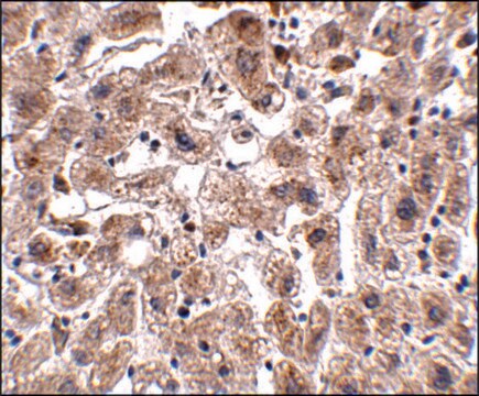
Immunohistochemistry Analysis: 1:500 dilution from a previous lot detected ZO-1 in normal human testis tissue.
Research Category
Neuroscience
Research Sub Category
Neuronal & Glial Markers

Quality

Evaluated by Western Blot in HepG2 cell lysate.

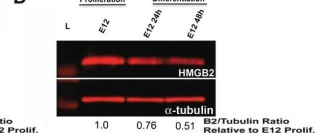
Western Blot Analysis: 0.1 µg/mL of this antibody detected ZO-1 on 10 µg of HepG2 cell lysate.

Target description

~260 kDa observed

Physical form

Affinity purified
Purified rabbit polyclonal in buffer containing 0.1 M Tris-Glycine (pH 7.4), 150 mM NaCl with 0.05% sodium azide.

Storage and Stability

Stable for 1 year at 2-8°C from date of receipt.

Analysis Note

Control
HepG2 cell lysate

Other Notes

Concentration: Please refer to lot specific datasheet.
Concentration: Please refer to the Certificate of Analysis for the lot-specific concentration.

Disclaimer

Unless otherwise stated in our catalog or other company documentation accompanying the product(s), our products are intended for research use only and are not to be used for any other purpose, which includes but is not limited to, unauthorized commercial uses, in vitro diagnostic uses, ex vivo or in vivo therapeutic uses or any type of consumption or application to humans or animals.

Not finding the right product?  

Try our Product Selector Tool.

Storage Class Code

12 - Non Combustible Liquids

WGK

WGK 1

Flash Point(F)

Not applicable

Flash Point(C)

Not applicable


Certificates of Analysis (COA)

Search for Certificates of Analysis (COA) by entering the products Lot/Batch Number. Lot and Batch Numbers can be found on a product’s label following the words ‘Lot’ or ‘Batch’.

Already Own This Product?

Find documentation for the products that you have recently purchased in the Document Library.

Visit the Document Library

Lin Zhu et al.
Atherosclerosis, 268, 117-126 (2017-12-05)
Gut microbiota plays a major role in metabolic disorders. Berberine is used to treat obesity, diabetes and atherosclerosis. The mechanism underlying the role of berberine in modulating metabolic disorders is not fully clear because berberine has poor oral bioavailability. Thus
Mikolaj Adamek et al.
Virulence, 12(1), 1855-1883 (2021-07-17)
Gills of fish are involved in respiration, excretion and osmoregulation. Due to numerous interactions between these processes, branchial diseases have serious implications on fish health. Here, "koi sleepy disease" (KSD), caused by carp edema virus (CEV) infection was used to
Leopoldina Scotti et al.
Reproduction, fertility, and development, 28(6), 690-699 (2014-12-09)
Ovarian hyperstimulation syndrome (OHSS) is a complication of ovarian stimulation with gonadotrophins following human chorionic gonadotrophin (hCG) administration. The relationship between hCG and OHSS is partly mediated via the production of angiogenic factors, such as vascular endothelial growth factor A
Dina Gadalla et al.
Journal of biomedical materials research. Part A, 110(6), 1251-1262 (2022-02-11)
There are reciprocal interactions between epithelial cells and underlying basement membrane. The resemblance of biomaterials to native basement membrane is thus critical for their success when used to regenerate epithelium-containing organs. Particularly, the use of nanofibers and the incorporation of
Sung-Eun Choi et al.
Experimental and therapeutic medicine, 24(5), 648-648 (2022-09-29)
Tubulointerstitial fibrosis is characterized by accumulation of the extracellular matrix in the interstitium. Lysyl oxidase-like 2 (LOXL2), a member of the lysyl oxidase family, is known for promoting cancer metastasis, invasion and stromal fibrosis in various organs. Our previous study

Articles

16HBE14o- human bronchial epithelial cells used to model respiratory epithelium for the research of cystic fibrosis, viral pulmonary pathology (SARS-CoV), asthma, COPD, effects of smoking and air pollution. See over 5k publications.

16HBE14o- human bronchial epithelial cells used to model respiratory epithelium for the research of cystic fibrosis, viral pulmonary pathology (SARS-CoV), asthma, COPD, effects of smoking and air pollution. See over 5k publications.

16HBE14o- human bronchial epithelial cells used to model respiratory epithelium for the research of cystic fibrosis, viral pulmonary pathology (SARS-CoV), asthma, COPD, effects of smoking and air pollution. See over 5k publications.

16HBE14o- human bronchial epithelial cells used to model respiratory epithelium for the research of cystic fibrosis, viral pulmonary pathology (SARS-CoV), asthma, COPD, effects of smoking and air pollution. See over 5k publications.

Our team of scientists has experience in all areas of research including Life Science, Material Science, Chemical Synthesis, Chromatography, Analytical and many others.

Contact Technical Service