Monoclonal Anti-τ (Tau) antibody produced in mouse, clone TAU-2, ascites fluid
biological source
mouse
conjugate
unconjugated
antibody form
ascites fluid
antibody product type
primary antibodies
clone
TAU-2, monoclonal
mol wt
antigen 55-62 kDa
contains
15 mM sodium azide
species reactivity
monkey, bovine, chicken, human
technique(s)
immunohistochemistry (formalin-fixed, paraffin-embedded sections): suitable microarray: suitable western blot: 1:1,000 using a fresh total bovine brain extract or an enriched microtubule protein preparation
Monoclonal Anti- τ (TAU) (mouse IgG1 isotype) is derived from the hybridoma produced by the fusion of mouse myeloma cells and splenocytes from an immunized mouse. τ (TAU) proteins are a part of microtubule associated proteins (MAPs). They are densely found in neurons and in trace amounts in non-neuronal cells. In brain six isoforms of τ (TAU) proteins are present.
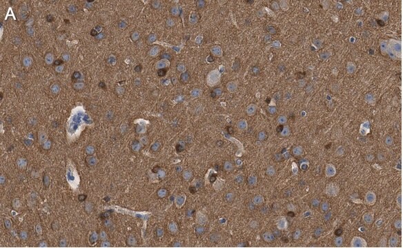
The antibody reacts exclusively with the chemically heterogeneous τ in both the phosphorylated and non-phosphorylated form. The antibody does not react with other MAPs or with tubulin. In immunohistochemical staining, it localizes τ along microtubules in axons, somata, dendrites and astrocytes, and on ribosomes. The antibody may be used for staining of τ in Alzheimer neurofibrillary tangles in sections of human brain tissue.
The best known microtubule associated proteins (MAPs) which copurify with microtubules are MAP2 and Tau. These two proteins are heat stable and stimulate formation of the microtubule polymer from purified tubulin subunits. Tau is chemically heterogenous, however, limited protolysis has demonstrated that the different eletrophoretic species are closely related. Tau is immunologically distinct from the other MAPs, namely MAP1, MAP2 and MAP5. Localization studies have demonstrated that Tau is intimately associated with the filamentous structures which compose the neurofibrillary tangles as found in an Alzheimer′s disease brain.
Immunogen
bovine microtubule-associated proteins (MAPs)
Application
Monoclonal Anti-τ (Tau) antibody has been used:
in immunohistology
in immunoblotting
in dot blot
in immunohistochemistry
Mouse monoclonal clone TAU-2 anti-Tau antibody maybe used to study microtubule associated proteins (MAP) expression and cytological localization in various tissue and cell lines, under different developmental and environmental circumstances.
Biochem/physiol Actions
Monoclonal Anti- τ (TAU) is phosphatase independent; it will bind Tau proteins in either their phosphorylated or non-phosphorylated forms. It localizes Tau proteins along microtubules in axons, somata, dendrites, astrocytes and on ribosomes (polysomes). The best-known microtubule associated proteins (MAPs) which copurify with microtubules are MAP2 and Tau. These two proteins are heat stable and stimulate formation of the microtubule polymer from purified tubulin subunits. Tau is immunologically distinct from the other MAPs. Tau is intimately associated with the filamentous structures which compose the neurofibrillary tangles as found in an Alzheimer′s disease brain.
Disclaimer
Unless otherwise stated in our catalog or other company documentation accompanying the product(s), our products are intended for research use only and are not to be used for any other purpose, which includes but is not limited to, unauthorized commercial uses, in vitro diagnostic uses, ex vivo or in vivo therapeutic uses or any type of consumption or application to humans or animals.
International journal of molecular sciences, 23(1) (2022-01-12)
Alzheimer's disease (AD) is the leading cause of dementia among the elderly. Neuropathologically, AD is characterized by the deposition of a 39- to 42-amino acid long β-amyloid (Aβ) peptide in the form of senile plaques. Several post-translational modifications (PTMs) in
Presenilin-1 (PS-1) mutations can cause Pick's disease without evidence of Alzheimer's disease (AD). We describe a family with a PS-1 M146L mutation and both Pick bodies and AD. Sarkosyl-insoluble hyperphosphorylated tau showed three bands consistent with AD, although dephosphorylation showed
Brain : a journal of neurology, 127(Pt 10), 2214-2220 (2004-07-30)
The main unifying feature of cases with frontotemporal dementia (FTD) is the pattern of brain atrophy. Surprisingly, there are a variety of underlying histopathologies in cases with the clinical features and typical pattern of atrophy characterizing FTD. This suggests that
Cortical limb myoclonus in pathologically proven progressive supranuclear palsy.
Sharon Kemp et al.
Movement disorders : official journal of the Movement Disorder Society, 28(13), 1804-1806 (2013-10-15)
Journal of molecular neuroscience : MN, 45(1), 32-41 (2011-02-23)
Lesions containing aggregated and hyperphosphorylated tau protein are characteristic of neurodegenerative tauopathies. We have developed a cellular model of pathological tau deposition and clearance by overexpressing wild type human tau in HEK293 cells. When proteasome activity is inhibited, HEK293/tau cells
Our team of scientists has experience in all areas of research including Life Science, Material Science, Chemical Synthesis, Chromatography, Analytical and many others.