Skip to Content
Merck
All Photos(1)

Documents

408R-1

Sigma-Aldrich

S100A1 (EP184) Rabbit Monoclonal Primary Antibody

Sign Into View Organizational & Contract Pricing


About This Item

UNSPSC Code:
12352203
NACRES:
NA.41

biological source

rabbit

Quality Level

100
500

conjugate

unconjugated

antibody form

culture supernatant

antibody product type

primary antibodies

clone

EP184, monoclonal

description

For In Vitro Diagnostic Use in Select Regions (See Chart)

form

buffered aqueous solution

species reactivity

human

packaging

vial of 0.1 mL concentrate (408R-14)
vial of 0.5 mL concentrate (408R-15)
bottle of 1.0 mL predilute (408R-17)
vial of 1.0 mL concentrate (408R-16)
bottle of 7.0 mL predilute (408R-18)

manufacturer/tradename

Cell Marque

technique(s)

immunohistochemistry (formalin-fixed, paraffin-embedded sections): 1:25-1:100

isotype

IgG

control

kidney, renal oncocytoma

shipped in

wet ice

storage temp.

2-8°C

visualization

cytoplasmic, nuclear

Gene Information

human ... S100A1(6271)

General description

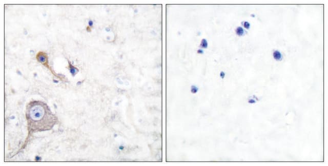
S100A1 is a member of S100 protein family of calcium binding proteins.S100A1 is very useful in the identification of renal cell carcinoma.Published literature indicates that 37 of 40 renal oncocytomas (93%), including two with the tubulo-cystic growth pattern, reacted positively by anti-S100A1.Most of them showed both cytoplasmic and nuclear immunostaining with moderate-to-strong intensity. S100A1 immunostaining was also observed in 30 of 41 clear cell renal cell carcinomas (73%); 6 showed focal positivity, whereas 24 displayed an immunoreactivity ranging from moderate (12 cases) to diffuse (12 cases) expression. Anti-S100A1 detected 30 cases of 32 papillary renal cell carcinomas (94%), 6 showing focal, 12 moderate, and 12 diffuse immunoreactivity. This study also demonstrated that 48 of the 51 chromophobe renal cell carcinomas (94%) showed no labeling by anti-S100A1. In 3 cases of chromophobe renal cell carcinomas (6%) that were positive by anti-S100A1, 2 were classic variant showing scattered S100A1 immunoreactive cells and an eosinophilic variant with nucleocytoplasmic staining in about 40% of the neoplastic cells.Therefore, anti-S100A1 is very useful in the differentiation of renal oncocytoma from chromophobe renal cell carcinoma as well as distinguishing clear cell renal cell carcinoma from chromophobe renal cell carcinoma. S100A1 is detected in normal renal tissue and expressed in both the nuclei and the cytoplasm of the cells lining proximal tubules, loops of Henle, and collecting ducts. The glomerular components are negative. S100A1 is also expressed by skeletal muscle and myocardium; and specific cytoplasmic expression is found in follicular dendritic cells of lymph node.

Quality


IVD

IVD

IVD

RUO

Linkage

S100A1 Positive Control Slides, Product No. 408S, are available for immunohistochemistry (formalin-fixed, paraffin-embedded sections).

Physical form

Solution in Tris Buffer, pH 7.3-7.7, with 1% BSA and <0.1% Sodium Azide

Preparation Note

Download the IFU specific to your product lot and formatNote: This requires a keycode which can be found on your packaging or product label.

Other Notes

For Technical Service please contact: 800-665-7284 or email: service@cellmarque.com

Legal Information

Cell Marque is a trademark of Merck KGaA, Darmstadt, Germany

Not finding the right product?  

Try our Product Selector Tool.

Storage Class Code

12 - Non Combustible Liquids

WGK

WGK 2

Flash Point(F)

Not applicable

Flash Point(C)

Not applicable


Certificates of Analysis (COA)

Search for Certificates of Analysis (COA) by entering the products Lot/Batch Number. Lot and Batch Numbers can be found on a product’s label following the words ‘Lot’ or ‘Batch’.

Already Own This Product?

Find documentation for the products that you have recently purchased in the Document Library.

Visit the Document Library

Paolo Cossu Rocca et al.
Modern pathology : an official journal of the United States and Canadian Academy of Pathology, Inc, 20(7), 722-728 (2007-05-08)
S100A1 is a calcium-binding protein, which has been recently found in renal cell neoplasms. We evaluated the diagnostic utility of immunohistochemical detection of S100A1 in 164 renal cell neoplasms. Forty-one clear cell, 32 papillary, and 51 chromophobe renal cell carcinomas
D B Zimmer et al.
Brain research bulletin, 37(4), 417-429 (1995-01-01)
The S100 family of calcium binding proteins contains approximately 16 members each of which exhibits a unique pattern of tissue/cell type specific expression. Although the distribution of these proteins is not restricted to the nervous system, the implication of several

Our team of scientists has experience in all areas of research including Life Science, Material Science, Chemical Synthesis, Chromatography, Analytical and many others.

Contact Technical Service