Skip to Content
Merck
All Photos(3)

Documents

MABT858

Sigma-Aldrich

Anti-prelamin A Antibody, clone PL-1C7

clone PL-1C7, from mouse

Synonym(s):

LMNA

Sign Into View Organizational & Contract Pricing


About This Item

UNSPSC Code:
12352203
eCl@ss:
32160702
NACRES:
NA.41

biological source

mouse

Quality Level

antibody form

purified antibody

antibody product type

primary antibodies

clone

PL-1C7, monoclonal

species reactivity

mouse, human

packaging

antibody small pack of 25 μg

technique(s)

ELISA: suitable
flow cytometry: suitable
immunocytochemistry: suitable
western blot: suitable

isotype

IgG2bκ

NCBI accession no.

UniProt accession no.

shipped in

ambient

target post-translational modification

unmodified

Gene Information

human ... LMNA(4000)

Related Categories

General description

Prelamin-A/C (UniProt: P02545; also known as 70 kDa lamin, Renal carcinoma antigen NY-REN-32) is encoded by the LMNA (also known as LMN1) gene (Gene ID: 4000) in human. Lamins are components of the nuclear lamina that provide a framework for the nuclear envelope and may also interact with chromatin. Lamin A and C are present in equal amounts in the lamina of mammals. Plays an important role in nuclear assembly, chromatin organization, nuclear membrane and telomere dynamics. Lamin A is initially synthesized as prelamin A that undergoes several modifications in the carboxyl terminal region that allow incorporation of prelamin A into the nuclear envelope and its subsequent processing into the mature lamin A. Cleavage of 15 residues (aa 647-662) by ZMPSTE24/FACE1 generates the final protein product. Unlike mature lamin A, prelamin A accumulates as discrete and localized foci at the nuclear periphery. Prelamin-A/C can accelerate smooth muscle cell senescence. It can act to disrupt mitosis and induce DNA damage in vascular smooth muscle cells (VSMCs), leading to mitotic failure, genomic instability, and premature senescence. Mutations in LMNA gene are known to cause Emery-Dreifuss muscular dystrophy that is characterized by weakness and atrophy of muscle without involvement of the nervous system. Some mutations have also been linked to familial type of lipodystrophy characterized by the loss of subcutaneous adipose tissue in the lower parts of the body. (Ref.: Casasola, A., et al. (2016). Nucleus 7(1); 84-102).

Specificity

Clone PL-1C7 detects Prelamin A in human and murine cells. It specifically recognizes prelamin A at the intact ZMPSTE24 cleavage site.

Immunogen

A lnear peptide corresponding to 12 amino acids from the C-terminal region of human Prelamin A.
Epitope: C-terminus

Application

Anti-prelamin A, clone PL-1C7, Cat. No. MABT858, is a mouse monoclonal antibody that detects Prelamin A and has been tested for use in ELISA, Flow Cytometry, Immunocytochemistry, and Western Blotting.
Research Category
Cell Structure
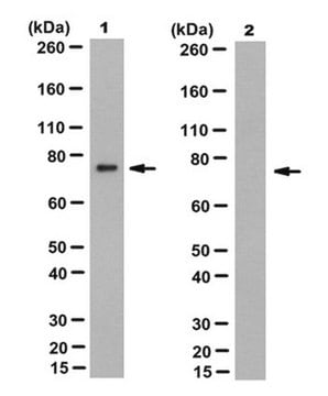
Western Blotting Analysis: 2 µg/mL from a representative lot detected prelamin A in U20S cells treated with farnesyl transferase inhibitor, Lonafarnib (3.2 uM for 24 h).

Flow Cytometry Analysis: A representative lot detected prelamin A in Flow Cytometry applications (Casasola, A., et. al. (2016). Nucleus. 7(1):84-102).

Western Blotting Analysis: A representative lot detected prelamin A in Western Blotting applications (Casasola, A., et. al. (2016). Nucleus. 7(1):84-102).

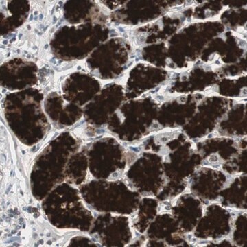
Immunocytochemistry Analysis: 1 µg/mL from a representative lot detected prelamin A in C2C12 cells with Farnesyl transferase inhibitor Lonafarnib (Courtesy of Fred Hutchinson Cancer Research Center, Seattle, Washington USA).

ELISA Analysis: A representative lot detected prelamin A in ELISA applications (Casasola, A., et. al. (2016). Nucleus. 7(1):84-102).

Immunocytochemistry Analysis: A representative lot detected prelamin A in Immunocytochemistry applications (Casasola, A., et. al. (2016). Nucleus. 7(1):84-102).

Quality

Evaluated by Western Blotting in C2C12 cell lysate.

Western Blotting Analysis: 2 µg/mL of this antibody detected prelamin A in C2C12 cell lysate.

Target description

~74 kDa observed; 74.14 kDa calculated. Uncharacterized bands may be observed in some lysate(s).

Physical form

Format: Purified
Protein G purified
Purified mouse monoclonal antibody IgG2b in buffer containing 0.1 M Tris-Glycine (pH 7.4), 150 mM NaCl with 0.05% sodium azide.

Storage and Stability

Stable for 1 year at 2-8°C from date of receipt.

Other Notes

Concentration: Please refer to lot specific datasheet.

Disclaimer

Unless otherwise stated in our catalog or other company documentation accompanying the product(s), our products are intended for research use only and are not to be used for any other purpose, which includes but is not limited to, unauthorized commercial uses, in vitro diagnostic uses, ex vivo or in vivo therapeutic uses or any type of consumption or application to humans or animals.

Not finding the right product?  

Try our Product Selector Tool.

Storage Class Code

12 - Non Combustible Liquids

WGK

WGK 1

Flash Point(F)

does not flash

Flash Point(C)

does not flash


Certificates of Analysis (COA)

Search for Certificates of Analysis (COA) by entering the products Lot/Batch Number. Lot and Batch Numbers can be found on a product’s label following the words ‘Lot’ or ‘Batch’.

Already Own This Product?

Find documentation for the products that you have recently purchased in the Document Library.

Visit the Document Library

Cristina Capanni et al.
Frontiers in cell and developmental biology, 10, 1018102-1018102 (2022-12-06)
Lamin A is a main constituent of the nuclear lamina and contributes to nuclear shaping, mechano-signaling transduction and gene regulation, thus affecting major cellular processes such as cell cycle progression and entry into senescence, cellular differentiation and stress response. The
Bo Jiang et al.
Science advances, 8(27), eabo0322-eabo0322 (2022-07-21)
Progerin, a product of LMNA mutation, leads to multiple nuclear abnormalities in patients with Hutchinson-Gilford progeria syndrome (HGPS), a devastating premature aging disorder. Progerin also accumulates during physiological aging. Here, we demonstrate that impaired insulin-like growth factor 1 receptor (IGF-1R)/Akt
Camilla Evangelisti et al.
Cells, 9(3) (2020-04-03)
A type lamins are fundamental components of the nuclear lamina. Changes in lamin A expression correlate with malignant transformation in several cancers. However, the role of lamin A has not been explored in osteosarcoma (OS). Here, we wanted to investigate
Francesca Chiarini et al.
Cell death & disease, 13(4), 346-346 (2022-04-16)
Lamin A, a main constituent of the nuclear lamina, is involved in mechanosignaling and cell migration through dynamic interactions with the LINC complex, formed by the nuclear envelope proteins SUN1, SUN2 and the nesprins. Here, we investigated lamin A role
Rouven Arnold et al.
International journal of molecular sciences, 22(14) (2021-07-25)
Hutchinson-Gilford progeria syndrome (HGPS) is an ultra-rare multisystem premature aging disorder that leads to early death (mean age of 14.7 years) due to myocardial infarction or stroke. Most cases have a de novo point mutation at position G608G within exon

Our team of scientists has experience in all areas of research including Life Science, Material Science, Chemical Synthesis, Chromatography, Analytical and many others.

Contact Technical Service