ABC1463
Ant-Zyxin
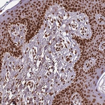
Anti-Zyxin Antibody, Cat. No. ABC1463 is a rabbit polyclonal antibody that detects Zyxin and has been tested for use in Immunocytochemistry and Western Blotting.
Synonym(s):
Zyxin-2
Sign Into View Organizational & Contract Pricing
All Photos(1)
About This Item
UNSPSC Code:
12352203
eCl@ss:
32160702
NACRES:
NA.43
Recommended Products
General description
Zyxin (UniProt: Q15942; also known as Zyxin-2) is encoded by the ZYX gene (Gene ID: 7791) in human. Zyxin is an evolutionarily conserved LIM domain protein that acts as an adhesion plaque protein concentrated at sites of cell adhesion and is widely expressed. It has been implicated in cell adhesion, cell motility, and signaling. Zyxin is reported to associate with members of the Enabled (Ena)/vasodilator-stimulated phosphoprotein (VASP) family of cytoskeletal regulators and plays a role in cytoskeletal dynamics and signaling. Zyxin contains three LIM zinc binding domains (aa 384-443; 444-503; and 504-570), which act as a negative regulator of Zyxin-VASP complexes. Also, it contains four proline rich ActA repeats (aa 64-77; 94-108; 115-121; and 127-137) that are known to interact directly with Ena/VASP proteins. Loss of Zyxin results in inability to thicken stress fibers in response to mechanical stimuli. (Ref.: Hoffman LM et al (2003). Mol. Cell Biol. 23(1); 70-79; Moody JD et al. (2009). Biochem. Biophys. Res. Commun. 378(3), 625-628; Yoshigi et al (2005). J. Cell Biol. 171(2), 209-215.).
Specificity
This rabbit polyclonal antibody detects Zyxin in human, mouse, and chicken cells. It targets an epitope within 20 amino acids from the C-terminal half.
Immunogen
Epitope: unknown
KLH-conjugated linear peptide corresponding to 20 amino acids from the C-terminal half of human Zyxin.
Application
Research Category
Cell Structure
Cell Structure
Western Blotting Analysis: A 1:1,000 dilution from a representative lot detected Zyxin in A431 cell lysate.
Immunofluorescence Analysis: A representative lot detected Zyxin in newborn mouse skin. Hoffman, L.M., et. al. (2003). Mol Cell Biol. 23(1):70-9; Hoffman, L.M., et. al. (2012). Mol Biol Cell. 23(10):1846-59).
Western Blotting Analysis): A representative lot detected Zyxin in mouse lung, mouse embryo fibroblasts and in human Ewing sarcoma cells.(Chaturvedi, A., et. al. (2014). Mol Biol Cell. 25(18):2695-709; Hoffman, L.M., et. al. (2003). Mol Cell Biol. 23(1):70-9; Hoffman, L.M., et. al. (2012). Mol Biol Cell. 23(10):1846-59; Hoffman, L.M., et. al. (2006). J Cell Biol. 172(5):771-82).
Immunocytochemistry Analysis: A representative lot detected Zyxin at focal adhesions in mouse fibroblasts and in 4T1 cells with ATG7 shRNA or ATG5 shRNA (Hoffman, L.M., et. al. (2006). J Cell Biol. 172(5):771-82; Sharifi, M.N., et. al. (2016). Cell Rep. 15(8):1660-72).
Quality
Evaluated by Western Blotting in NIH3T3 cell lysate.
Western Blotting Analysis: A 1:5,000 dilution of this antibody detected Zyxin in NIH3T3 cell lysate.
Western Blotting Analysis: A 1:5,000 dilution of this antibody detected Zyxin in NIH3T3 cell lysate.
Target description
~75 kDa observed; 61.28 kDa calculated. Uncharacterized bands may be observed in some lysate(s).
Physical form
Rabbit polyclonal antiserum with 0.05% sodium azide.
Unpurified
Storage and Stability
Stable for 1 year at -20°C from date of receipt.
Handling Recommendations: Upon receipt and prior to removing the cap, centrifuge the vial and gently mix the solution. Aliquot into microcentrifuge tubes and store at -20°C. Avoid repeated freeze/thaw cycles, which may damage IgG and affect product performance.
Handling Recommendations: Upon receipt and prior to removing the cap, centrifuge the vial and gently mix the solution. Aliquot into microcentrifuge tubes and store at -20°C. Avoid repeated freeze/thaw cycles, which may damage IgG and affect product performance.
Other Notes
Concentration: Please refer to lot specific datasheet.
Disclaimer
Unless otherwise stated in our catalog or other company documentation accompanying the product(s), our products are intended for research use only and are not to be used for any other purpose, which includes but is not limited to, unauthorized commercial uses, in vitro diagnostic uses, ex vivo or in vivo therapeutic uses or any type of consumption or application to humans or animals.
Certificates of Analysis (COA)
Search for Certificates of Analysis (COA) by entering the products Lot/Batch Number. Lot and Batch Numbers can be found on a product’s label following the words ‘Lot’ or ‘Batch’.
Already Own This Product?
Find documentation for the products that you have recently purchased in the Document Library.
Our team of scientists has experience in all areas of research including Life Science, Material Science, Chemical Synthesis, Chromatography, Analytical and many others.
Contact Technical Service


