Skip to Content
Merck
All Photos(1)

Documents

AB5585

Sigma-Aldrich

Anti-Recoverin Antibody

serum, Chemicon®

Synonym(s):

Recoverin

Sign Into View Organizational & Contract Pricing


About This Item

UNSPSC Code:
12352203
eCl@ss:
32160702
NACRES:
NA.41

biological source

rabbit

antibody form

serum

antibody product type

primary antibodies

clone

polyclonal

species reactivity

human, mouse, rat, frog, chicken

manufacturer/tradename

Chemicon®

technique(s)

immunocytochemistry: suitable
immunohistochemistry (formalin-fixed, paraffin-embedded sections): suitable
western blot: suitable

NCBI accession no.

UniProt accession no.

shipped in

dry ice

target post-translational modification

unmodified

Gene Information

human ... RCVRN(5957)
mouse ... Rcvrn(19674)

General description

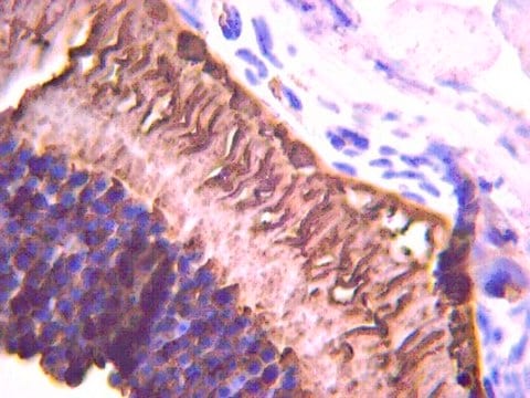
Recoverin is a calcium-binding protein expressed in retinal photoreceptors and midget cone bipolar cells. It appears to delay the termination of the phototransduction cascade by blocking the phosphorylation of photoexcited rhodopsin.

Specificity

Other species have not been tested.
Recognizes recoverin.

Immunogen

Recombinant human recoverin.

Application

Detect Recoverin using this Anti-Recoverin Antibody validated for use in IC, IH, IH(P) & WB.
Research Category
Neuroscience
Research Sub Category
Sensory & PNS
Western Blot Analysis:
1:1,000-1:5,000 dilution using ECL.

Immunohistochemistry:
1:1,000 (2 hours at room temperature or overnight at 2-8ºC) on frozen and paraffin embedded tissue sections. Suggested fixative is 4% paraformaldehyde or 3% paraformaldehyde with 0.1% glutaraldehyde. Suggested permeabilization solution is 0.1% Triton X-100. Suggested blocking buffer is 0.05 M Tris saline, pH 7.4 containing 2.5% BSA, 0.1% triton and 0.05% sodium azide.

Optimal working dilutions must be determined by the end user.

Target description

26 kDa

Linkage

Replaces: AB5431P

Physical form

Rabbit polyclonal serum. Contains no preservative.
Unpurified

Storage and Stability

Stable for 1 year at -20ºC from date of receipt.

Analysis Note

Control
POSITIVE CONTROL: Recoverin is found in photoreceptors and midget cone bipolar cells in retina and pinealocytes in pineal gland.

Other Notes

Concentration: Please refer to the Certificate of Analysis for the lot-specific concentration.

Legal Information

CHEMICON is a registered trademark of Merck KGaA, Darmstadt, Germany

Disclaimer

Unless otherwise stated in our catalog or other company documentation accompanying the product(s), our products are intended for research use only and are not to be used for any other purpose, which includes but is not limited to, unauthorized commercial uses, in vitro diagnostic uses, ex vivo or in vivo therapeutic uses or any type of consumption or application to humans or animals.

Not finding the right product?  

Try our Product Selector Tool.

Storage Class Code

11 - Combustible Solids

WGK

WGK 1

Flash Point(F)

Not applicable

Flash Point(C)

Not applicable


Certificates of Analysis (COA)

Search for Certificates of Analysis (COA) by entering the products Lot/Batch Number. Lot and Batch Numbers can be found on a product’s label following the words ‘Lot’ or ‘Batch’.

Already Own This Product?

Find documentation for the products that you have recently purchased in the Document Library.

Visit the Document Library

Katharina Kruszewski et al.
Investigative ophthalmology & visual science, 57(3), 1120-1131 (2016-03-15)
Retinal degeneration is a common feature of several lysosomal storage disorders, including the mucopolysaccharidoses, a group of metabolic disorders that is characterized by widespread accumulation of glycosaminoglycans due to lysosomal enzyme dysfunction. We used a new mouse model of mucopolysaccharidosis
Kent W Small et al.
Ophthalmology, 123(1), 9-18 (2015-10-29)
To identify specific mutations causing North Carolina macular dystrophy (NCMD). Whole-genome sequencing coupled with reverse transcription polymerase chain reaction (RT-PCR) analysis of gene expression in human retinal cells. A total of 141 members of 12 families with NCMD and 261
Sonia Biswas et al.
PloS one, 9(6), e99075-e99075 (2014-06-12)
In the widely used mouse model of retinal degeneration, rd1, the loss of photoreceptors leads to rhythmic electrical activity of around 10-16 Hz in the remaining retinal network. Recent studies suggest that this oscillation is formed within the electrically coupled
J Cui et al.
Neuroscience, 224, 63-69 (2012-08-23)
Retinal bipolar cells and ganglion cells are known to possess voltage-gated T-type Ca(2+) channels. Previous electrophysiological recording studies suggested that there is differential expression of different T-type Ca(2+) channel α1 subunits among bipolar cells. The detailed expression patterns of the
Amani A Fawzi et al.
Amyotrophic lateral sclerosis & frontotemporal degeneration, 15(7-8), 569-580 (2014-10-17)
Our objective was to present clinicopathologic evidence of anterior visual pathway involvement in patients with amyotrophic lateral sclerosis (ALS) secondary to a C9orf72 mutation. Two related patients from an extended pedigree with ALS and GGGGCC hexanucleotide repeat expansion in the

Our team of scientists has experience in all areas of research including Life Science, Material Science, Chemical Synthesis, Chromatography, Analytical and many others.

Contact Technical Service