Skip to Content
Merck
All Photos(1)

Key Documents

B7401

Sigma-Aldrich

Anti-Mouse IgG (Fc specific)–Biotin antibody produced in goat

affinity isolated antibody, buffered aqueous solution

Synonym(s):

Goat Anti-Mouse IgG

Sign Into View Organizational & Contract Pricing


About This Item

MDL number:
UNSPSC Code:
12352203
NACRES:
NA.46

biological source

goat

conjugate

biotin conjugate

antibody form

affinity isolated antibody

antibody product type

secondary antibodies

clone

polyclonal

form

buffered aqueous solution

technique(s)

direct ELISA: 1:160,000
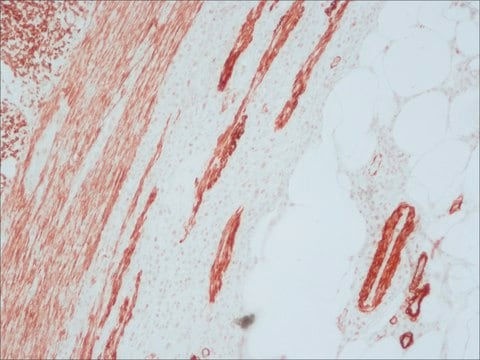
immunohistochemistry (formalin-fixed, paraffin-embedded sections): 1:400
western blot (chemiluminescent): 1:300,000 using total cell extract of HeLa cells (5-10 μg/well)

storage temp.

−20°C

target post-translational modification

unmodified

Looking for similar products? Visit Product Comparison Guide

General description

Goat polyclonal anti-Mouse IgG (Fc specific)–Biotin reacts with mouse IgG and mouse IgG, Fc fragment. It shows no reactivity with mouse IgG Fab fragment, human IgG, IgA, IgM, kappa and lambda light chain, bovine IgG and IgM, or horse IgG.
Mouse IgG is a plasma B cell derived antibody isotype defined by its heavy chain. IgG is the most abundant antibody isotype found in mouse serum. IgG crosses the placental barrier, is a complement activator and binds to the Fc-receptors on phagocytic cells. The level of IgG may vary with the status of disease or infection.
Biotin:avidin second antibody signal systems involve the conjugation of biotin to a secondary antibody. Once the secondary antibody binds to a target primary antibody in an immunochemical assay, the signal is developed by allowing avidin which has been conjugated to an enzyme or signal molecule to bind to the antibody associated biotin molecule.

Application

Anti-Mouse IgG (Fc specific)−Biotin antibody produced in goat has been used in ELISA and quantitative suspension array technology (qSAT) multiplex antibody assay.
Goat polyclonal anti-Mouse IgG (Fc specific)–Biotin may be used to detect mouse IgG within serum or other biological systems. The antibody may be used as a secondary antibody in immunochemical assays based on biotin:avidin second antibody signal systems wherein mouse IgG is the primary antibody.

Other Notes

Antibody adsorbed with bovine, equine and human serum proteins.

Physical form

Solution in 0.01 M phosphate buffered saline, pH 7.4, containing 15 mM sodium azide.

Preparation Note

Adsorbed to reduce background staining with bovine, horse or human samples.

Disclaimer

Unless otherwise stated in our catalog or other company documentation accompanying the product(s), our products are intended for research use only and are not to be used for any other purpose, which includes but is not limited to, unauthorized commercial uses, in vitro diagnostic uses, ex vivo or in vivo therapeutic uses or any type of consumption or application to humans or animals.

Not finding the right product?  

Try our Product Selector Tool.

Storage Class Code

12 - Non Combustible Liquids

WGK

nwg

Flash Point(F)

Not applicable

Flash Point(C)

Not applicable


Certificates of Analysis (COA)

Search for Certificates of Analysis (COA) by entering the products Lot/Batch Number. Lot and Batch Numbers can be found on a product’s label following the words ‘Lot’ or ‘Batch’.

Already Own This Product?

Find documentation for the products that you have recently purchased in the Document Library.

Visit the Document Library

The context of tetanus toxoid application influences the outcome of antigen-specific and self-directed humoral immune response
Stojanovic M, et al.
Microbiology and Immunology, 53(2), 89-100 (2009)
M B Merriman et al.
Clinical & experimental ophthalmology, 29(4), 248-252 (2001-09-08)
5-Fluorouracil (5-FU) is used intraoperatively to improve the success of trabeculectomy Common practice is a 5-min application of 5-FU delivered via a Weck Cel sponge. The aim of this study was to determine if shorter exposure times were as effective
Masahiro Kozuka et al.
Cancers, 13(19) (2021-10-14)
Circulating tumor cells (CTCs) are a prognostic marker in patients with metastatic colorectal cancer (mCRC). However, little is known about the characterization of CTCs in mCRC at the single-cell level using RNA sequencing. The purpose of this study was to
Mauro Corrado et al.
Cell death and differentiation, 28(7), 2194-2206 (2021-03-03)
Optic atrophy 1 (OPA1), a mitochondria-shaping protein controlling cristae biogenesis and respiration, is required for memory T cell function, but whether it affects intrathymic T cell development is unknown. Here we show that OPA1 is necessary for thymocyte maturation at
Bira Arumndari Nurrahma et al.
Frontiers in aging neuroscience, 13, 668775-668775 (2021-05-25)
Parkinson's disease (PD) is a neurodegenerative disease associated with progressive impairment of motor and non-motor functions in aging people. Overwhelming evidence indicate that mitochondrial dysfunction is a central factor in PD pathophysiology, which impairs energy metabolism. While, several other studies

Our team of scientists has experience in all areas of research including Life Science, Material Science, Chemical Synthesis, Chromatography, Analytical and many others.

Contact Technical Service