Skip to Content
Merck
All Photos(5)

Key Documents

07-1401

Sigma-Aldrich

Anti-GLUT-1 Antibody, CT

from rabbit, purified by affinity chromatography

Synonym(s):

Glucose Transporter type 1, Solute carrier family 2, facilitated glucose transporter, member 1

Sign Into View Organizational & Contract Pricing


About This Item

UNSPSC Code:
12352203
eCl@ss:
32160702
NACRES:
NA.41

biological source

rabbit

Quality Level

antibody form

purified immunoglobulin

antibody product type

primary antibodies

clone

polyclonal

purified by

affinity chromatography

species reactivity

human

species reactivity (predicted by homology)

mouse (Human. Predicted to react with Mouse based on 100% sequence homology.)

technique(s)

immunocytochemistry: suitable
immunohistochemistry: suitable (paraffin)
western blot: suitable

isotype

IgG

NCBI accession no.

UniProt accession no.

shipped in

wet ice

target post-translational modification

unmodified

Gene Information

human ... SLC2A1(6513)

General description

Solute carrier family 2, facilitated glucose transporter member 1 (UniProt: P11166; also known as Glucose transporter type 1, erythrocyte/brain, GLUT-1, HepG2 glucose transporter) is encoded by the SLC2A1 (also known as GLUT1) gene (Gene ID: 6513) in human. Glucose transporters are a family of integral membrane proteins that facilitative glucose uptake by cells. Seven different glucose transport proteins have been described that are designated as GLUT-1 to 7. GLUT-1 is a highly conserved; ubiquitously distributed, multi-pass membrane protein that is responsible for constitutive glucose uptake. It displays very broad substrate specificity and can transport a wide range of aldoses including both pentoses and hexoses. It is the predominant glucose transporter in embryonic and fetal tissues. In many organs, GLUT-1 is concentrated in endothelial cells of blood-tissue barriers. Hence, it has a specialized role to shuttle glucose between blood and organs that have limited access to small solutes via passive diffusion. It is also abundant in the mammalian erythrocyte membrane where it can rapidly equilibrate glucose between the cytoplasm of the erythrocyte and the blood plasma. GLUT-1 levels are reported to be frequently upregulated during tumorigenesis. Mutations in SLC2A1 gene are linked to GLUT-1 deficiency syndrome 1 and 2 that are characterized by infantile-onset epileptic encephalopathy, delayed development, microcephaly, and motor incoordination, and paroxysmal exercise-induced dyskinesia.

Specificity

This rabbit polyclonal antibody specifically detects Glucose transporter type 1 (GLUT-1). It targets an epitope within 12 amino acids from the C-terminal region.

Immunogen

Epitope: C-Terminus
KLH-conjugated linear peptide corresponding to the C-terminal of Human Glucose transporter-1 (GLUT-1).

Application

Anti-GLUT-1, CT, Cat. No. 07-1401, is a rabbit polyclonal antibody that detects glucose transporter member 1 and is tested for use in Western Blotting, Immunohistochemistry (Paraffin), and Immunocytochemistry
Research Category
Signaling
Research Sub Category
Insulin/Energy Signaling
Tested Applications
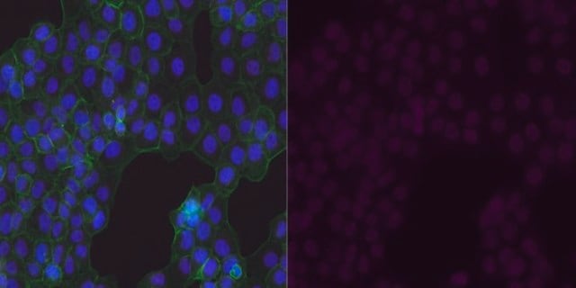
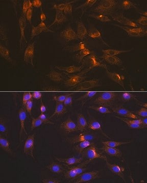
  • Immunocytochemistry Analysis: A 1:500 dilution from a representative lot detected GLUT-1 in A431 cells.
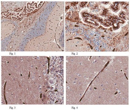
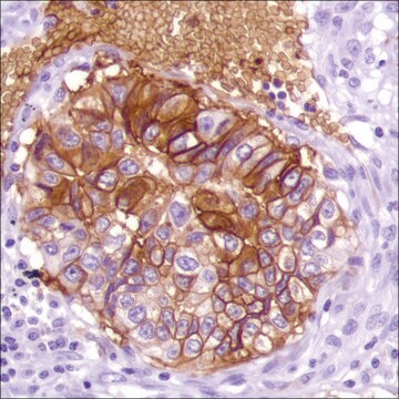
  • Immunohistochemistry (Paraffin) Analysis: A 1:1,000 dilution from a representative lot detected GLUT-1 in Human pancreas, Human lung, and Human placenta tissue sections.
  • Note: Actual optimal working dilutions must be determined by end user as specimens, and experimental conditions may vary with the end user.

Quality

Evaluated by Western Blotting in Human umbilical vein endothelial cell (HUVEC) lysate.
  • Western Blotting Analysis: A 1:1,000 dilution of this antibody detected GLUT-1 in Human umbilical vein endothelial cell (HUVEC) lysate.

Target description

~54 kDa observed; 54.08 kDa calculated. Uncharacterized bands may be observed in some lysate(s).

Linkage

Replaces: AB1340

Physical form

Affinity purified
Purified rabbit polyclonal antibody in buffer containing 0.02 M phosphate buffer, pH 7.6, 0.25 M NaCl, and 0.1% sodium azide.

Storage and Stability

Recommended storage: +2°C to +8°C.

Analysis Note

Control
Jurkat Cell Lysate

Other Notes

Concentration: Please refer to the Certificate of Analysis for the lot-specific concentration.

Disclaimer

Unless otherwise stated in our catalog or other company documentation accompanying the product(s), our products are intended for research use only and are not to be used for any other purpose, which includes but is not limited to, unauthorized commercial uses, in vitro diagnostic uses, ex vivo or in vivo therapeutic uses or any type of consumption or application to humans or animals.

Not finding the right product?  

Try our Product Selector Tool.

Storage Class Code

12 - Non Combustible Liquids

WGK

WGK 2

Flash Point(F)

Not applicable

Flash Point(C)

Not applicable


Certificates of Analysis (COA)

Search for Certificates of Analysis (COA) by entering the products Lot/Batch Number. Lot and Batch Numbers can be found on a product’s label following the words ‘Lot’ or ‘Batch’.

Already Own This Product?

Find documentation for the products that you have recently purchased in the Document Library.

Visit the Document Library

GLUT1 protein expression correlates with unfavourable histologic category and high risk in patients with neuroblastic tumours.
Ramani, Pramila, et al.
Virchows Archiv (2013)
Sugar transporter regulation by ATP and quaternary structure.
Cloherty, E K, et al.
Blood Cells, Molecules and Diseases, 27, 102-107 (2001)
Expression of glucose transporter GLUT3 by endotoxin in cultured rat astrocytes: the role of nitric oxide.
Cidad, P, et al.
Journal of Neurochemistry, 79, 17-24 (2001)
I W Smoak et al.
Anatomy and embryology, 201(5), 327-333 (2000-06-06)
The embryonic heart depends on glucose during early organogenesis. Glut-1 functions in constitutive glucose uptake in adult tissues and is the predominant glucose transporter in embryonic and fetal tissues. This study focuses on Glut-1 expression in the heart during normal
Critical role for lactate dehydrogenase A in aerobic glycolysis that sustains pulmonary microvascular endothelial cell proliferation.
Parra-Bonilla, G; Alvarez, DF; Al-Mehdi, AB; Alexeyev, M; Stevens, T
American Journal of Physiology. Lung Cellular and Molecular Physiology null

Our team of scientists has experience in all areas of research including Life Science, Material Science, Chemical Synthesis, Chromatography, Analytical and many others.

Contact Technical Service