Skip to Content
Merck
All Photos(2)

Key Documents

05-163

Sigma-Aldrich

Anti-PLCγ-1 Antibody

Upstate®, from mouse

Synonym(s):

1-phosphatidylinositol-4,5-bisphosphate phosphodiesterase gamma 1, Phosphoinositide phospholipase C, Phospholipase C-gamma-1, monophosphatidylinositol phosphodiesterase, phosphatidylinositol phospholipase C, phosphoinositidase C, phospholipase C gamma 1,

Sign Into View Organizational & Contract Pricing


About This Item

UNSPSC Code:
12352203
eCl@ss:
32160702
NACRES:
NA.41

biological source

mouse

Quality Level

antibody form

purified immunoglobulin

antibody product type

primary antibodies

clone

B-2-5, monoclonal
B-20-3, monoclonal
B-6-4, monoclonal
D-7-3, monoclonal
E-9-4, monoclonal

species reactivity

mouse, human, bovine, rat, rabbit

manufacturer/tradename

Upstate®

technique(s)

immunocytochemistry: suitable
immunoprecipitation (IP): suitable
western blot: suitable

isotype

IgG

NCBI accession no.

UniProt accession no.

shipped in

wet ice

target post-translational modification

unmodified

Gene Information

human ... PLCG1(5335)

General description

Phospholipase C gamma 1 is a tyrosine kinase substrate for many receptor and nonreceptor tyrosine kinases. Activation of the enzyme produces two second messenger molecules, inositol 1,4,5-trisphosphate and diacylglycerol, which provoke the mobilization of intracellular Ca2+ and activation of protein kinase C. Although the mammalian genome encodes 10 known phosphoinositide specific PLCs, only the gamma 1 and gamma 2 isoforms are regulated by tyrosine kinase activity. PLC beta isoform activity is controlled by heterotrimeric G protein coupled receptors, while the mechanism of regulation of PLCgamma activity is unknown.

Specificity

This antibody recognizes PLCγ-1, Mr 135 kDa. Reacts with some non-PLCγ-1 SH-3 containing proteins, including nck, Mr 47kDa (Park, D., 1992).

Immunogen

Five individual clones raised against purified soluble phospholipase C (PLC)γ-1 from bovine brain.

Application

Anti-PLCγ-1 Antibody detects level of PLCγ-1 & has been published & validated for use in IC, IP & WB.
Immunoprecipitation:
4 µg of a previous lot of antibody immunoprecipitated PLCγ-1 from non-stimulated A431 cell lysate, previous lots immunoprecipitated PLCγ-1 from rabbit brain cytosol preparation.

Immunocytochemistry:
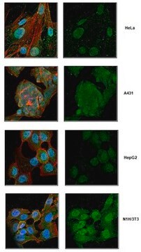
5-10 µg/mL of previous lots of this antibody detected PLCγ-1 on ethanol-acetic acid [95:5] fixed A431 cells.
Research Category
Signaling
Research Sub Category
Lipid Signaling

Quality

Routinely evaluated in human A431 carcinoma cells, rabbit brain cytosol, and bovine brain cytosol preparations.

Western Blot Analysis:
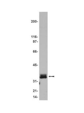
0.5-2 µg/mL of this lot detected PLCγ-1 in human A431 carcinoma cells; previous lots detected PKCγ-1 in rabbit brain cytosol and bovine brain cytosol preparations.

Target description

135 kDa

Physical form

Format: Purified
Liquid in 0.1 M Tris-glycine, pH 7.4, 0.15 M NaCl and 0.05% sodium azide.
An equal mix of IgG antibodies produced by SP2/0-Ag14 x Balb/c spleen cells (Suh, P.G., 1988) and propagated as purified immunoglobulin. Clones B-2-5, B-6-4, B-20-3, D-7-3, E-9-4.
Protein G Chromatography

Storage and Stability

Stable for 1 year at -20ºC from date of shipment.

Analysis Note

Control
Positive Antigen Control: Catalog #12-301, non-stimulated A431 cell lysate. Add 2.5µL of 2-mercaptoethanol/100µL of lysate and boil for 5 minutes to reduce the preparation. Load 20µg of reduced lysate per lane for minigels.

Legal Information

UPSTATE is a registered trademark of Merck KGaA, Darmstadt, Germany

Disclaimer

Unless otherwise stated in our catalog or other company documentation accompanying the product(s), our products are intended for research use only and are not to be used for any other purpose, which includes but is not limited to, unauthorized commercial uses, in vitro diagnostic uses, ex vivo or in vivo therapeutic uses or any type of consumption or application to humans or animals.

Not finding the right product?  

Try our Product Selector Tool.

Storage Class Code

12 - Non Combustible Liquids

WGK

WGK 1

Flash Point(F)

Not applicable

Flash Point(C)

Not applicable


Certificates of Analysis (COA)

Search for Certificates of Analysis (COA) by entering the products Lot/Batch Number. Lot and Batch Numbers can be found on a product’s label following the words ‘Lot’ or ‘Batch’.

Already Own This Product?

Find documentation for the products that you have recently purchased in the Document Library.

Visit the Document Library

J M Haugh et al.
The Journal of biological chemistry, 274(13), 8958-8965 (1999-03-20)
The epidermal growth factor receptor (EGFR) ligands, epidermal growth factor (EGF), and transforming growth factor-alpha (TGFalpha) elicit differential postendocytic processing of ligand and receptor molecules, which impacts long-term cell signaling outcomes. These differences arise from the higher affinity of the
A Zapf-Colby et al.
Oncogene, 18(35), 4908-4919 (1999-09-22)
Nerve growth factor (NGF) treatment of Chinese hamster ovary fibroblast (CHO) cells exogenously expressing 2.5x10⁵ TrkA receptors (CHO/TrkA) results in inhibition of serum and insulin-like growth factor-I (IGF-I) stimulated cell proliferation in a dose-dependent manner. Furthermore, NGF does not stimulate
L L Runft et al.
Developmental biology, 214(2), 399-411 (1999-10-20)
Elevation of intracellular Ca2+ at fertilization is essential for the initiation of development in the Xenopus egg, but the pathway between sperm-egg interaction and Ca2+ release from the egg's endoplasmic reticulum is not well understood. Here we show that injection
Yang Liu et al.
EBioMedicine, 51, 102607-102607 (2020-01-10)
PLCγ enzymes are key nodes in cellular signal transduction and their mutated and rare variants have been recently implicated in development of a range of diseases with unmet need including cancer, complex immune disorders, inflammation and neurodegenerative diseases. However, molecular
P M Irusta et al.
The EMBO journal, 17(23), 6912-6923 (1998-12-08)
Platelet-derived growth factor beta receptor (PDGFbetaR) is a transmembrane receptor tyrosine kinase involved in a variety of cellular functions. We have generated a constitutively activated murine PDGFbetaR containing a valine to alanine substitution at residue 536, located in the cytoplasmic

Our team of scientists has experience in all areas of research including Life Science, Material Science, Chemical Synthesis, Chromatography, Analytical and many others.

Contact Technical Service