
HPA038299
Anti-TMEM165 antibody produced in rabbit
Prestige Antibodies® Powered by Atlas Antibodies, affinity isolated antibody, buffered aqueous glycerol solution
Sinónimos:
Anti-GDT1, Anti-TMPT27, Anti-TPARL
About This Item
Productos recomendados


origen biológico
rabbit
conjugado
unconjugated
forma del anticuerpo
affinity isolated antibody
tipo de anticuerpo
primary antibodies
clon
polyclonal
Línea del producto
Prestige Antibodies® Powered by Atlas Antibodies
Formulario
buffered aqueous glycerol solution
reactividad de especies
human
técnicas
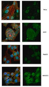
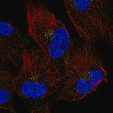
immunofluorescence: 0.25-2 μg/mL
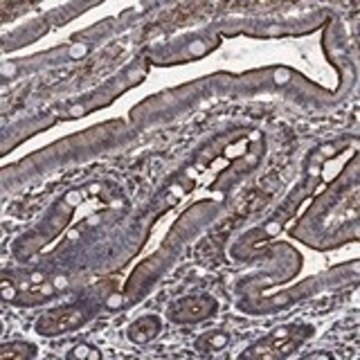
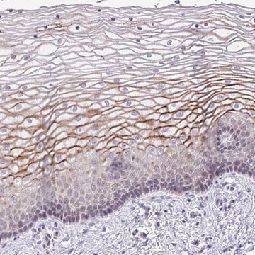
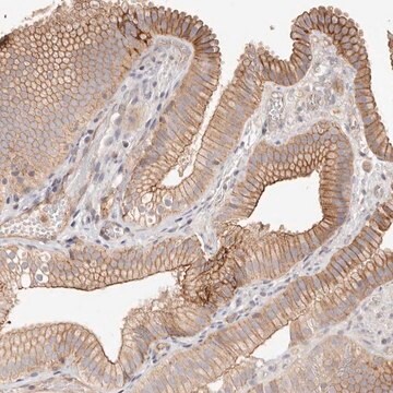
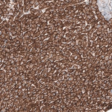
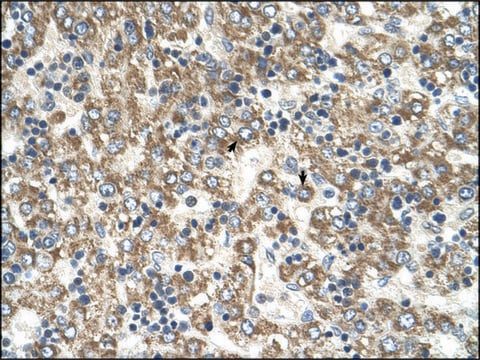
immunohistochemistry: 1:1000-1:2500
secuencia del inmunógeno
REGLKMSPDEGQEELEEVQAELKKKDEEFQRTKLLNGPGDVETGTSITVPQ
Nº de acceso UniProt
Condiciones de envío
wet ice
temp. de almacenamiento
−20°C
modificación del objetivo postraduccional
unmodified
Información sobre el gen
human ... TMEM165(55858)
Inmunógeno
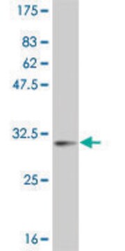
Aplicación
The Human Protein Atlas project can be subdivided into three efforts: Human Tissue Atlas, Cancer Atlas, and Human Cell Atlas. The antibodies that have been generated in support of the Tissue and Cancer Atlas projects have been tested by immunohistochemistry against hundreds of normal and disease tissues and through the recent efforts of the Human Cell Atlas project, many have been characterized by immunofluorescence to map the human proteome not only at the tissue level but now at the subcellular level. These images and the collection of this vast data set can be viewed on the Human Protein Atlas (HPA) site by clicking on the Image Gallery link. We also provide Prestige Antibodies® protocols and other useful information.
Características y beneficios
Every Prestige Antibody is tested in the following ways:
- IHC tissue array of 44 normal human tissues and 20 of the most common cancer type tissues.
- Protein array of 364 human recombinant protein fragments.
Ligadura / enlace
Forma física
Información legal
Cláusula de descargo de responsabilidad
¿No encuentra el producto adecuado?
Pruebe nuestro Herramienta de selección de productos.
Código de clase de almacenamiento
10 - Combustible liquids
Clase de riesgo para el agua (WGK)
WGK 1
Punto de inflamabilidad (°F)
Not applicable
Punto de inflamabilidad (°C)
Not applicable
Elija entre una de las versiones más recientes:
Certificados de análisis (COA)
¿No ve la versión correcta?
Si necesita una versión concreta, puede buscar un certificado específico por el número de lote.
¿Ya tiene este producto?
Encuentre la documentación para los productos que ha comprado recientemente en la Biblioteca de documentos.
Nuestro equipo de científicos tiene experiencia en todas las áreas de investigación: Ciencias de la vida, Ciencia de los materiales, Síntesis química, Cromatografía, Analítica y muchas otras.
Póngase en contacto con el Servicio técnico