Skip to Content
Merck
All Photos(2)

Key Documents

P6834

Sigma-Aldrich

Monoclonal Anti-Proliferating Cell Protein Ki-67 antibody produced in mouse

clone PP-67, ascites fluid

Synonym(s):

Anti-Ki-67

Sign Into View Organizational & Contract Pricing


About This Item

MDL number:
UNSPSC Code:
12352203
NACRES:
NA.41
conjugate:
unconjugated
application:
ARR
IHC (p)
clone:
PP-67, monoclonal
species reactivity:
human
citations:
27
technique(s):
immunoblotting: suitable
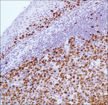
immunohistochemistry (formalin-fixed, paraffin-embedded sections): 1:800 using microwave-treated human tonsil sections
microarray: suitable

biological source

mouse

Quality Level

conjugate

unconjugated

antibody form

ascites fluid

antibody product type

primary antibodies

clone

PP-67, monoclonal

contains

15 mM sodium azide

species reactivity

human

technique(s)

immunoblotting: suitable
immunohistochemistry (formalin-fixed, paraffin-embedded sections): 1:800 using microwave-treated human tonsil sections
microarray: suitable

isotype

IgM

UniProt accession no.

shipped in

dry ice

storage temp.

−20°C

target post-translational modification

unmodified

Gene Information

human ... MKI67(4288)

General description

Marker of proliferation Ki-67 (MKI67) consists of a phosphopeptide-binding forkhead-associated (FHA) domain and a protein phosphatase 1 (PP1)-binding site at the amino- terminal, 16 tandem repeats at the central region and leucine and arginine (LR) pairs at the carboxy- terminal. It is expressed at all active phases of the cell cycle. The gene encoding this protein is localized on human chromosome 10.
Marker of proliferation Ki-67 (MKI67- antigen identified by monoclonal antibody) consists of a phosphopeptide-binding forkhead-associated (FHA) domain and a protein phosphatase 1 (PP1)-binding site at the amino- terminal, 16 tandem repeats at the central region and leucine and arginine (LR) pairs at the carboxy- terminal. It is expressed at all active phases (G1, S, G2, and mitosis) of the cell cycle. The gene encoding this protein is localized on human chromosome 10.

Specificity

Reacts with the Ki-67 nuclear antigen (345 and 395 kDa, and several lower m.w. proteins) in immunoblotting. The product shows an immunohistochemical staining pattern similar to that obtained with other anti-Ki-67/MIB-1 reagents. It labels proliferating cells in frozen human tissue and in formalin-fixed, paraffin-embedded tissue. A microwave heating retrieval step is required for successful labeling of routinely formalin-fixed, paraffin-embedded human tissue.

Immunogen

HeLa cell nuclear preparation.

Application

Applications in which this antibody has been used successfully, and the associated peer-reviewed papers, are given below.
Immunocytochemistry (1 paper)
Applications in which this antibody has been used successfully, and the associated peer-reviewed papers, are given below.
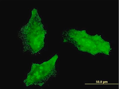
Immunocytochemistry.
Monoclonal Anti-Proliferating Cell Protein Ki-67 antibody produced in mouse has also been used for immunohistochemistry.

Biochem/physiol Actions

Marker of proliferation Ki-67 (MKI67), which is a part of the mitotic chromosome periphery, acts as a biological surfactant to maintain individual mitotic chromosomes dispersed in the cytoplasm after nuclear envelope disassembly. It is a prognostic marker in breast and prostate cancer. MKI67 is also a potential cell proliferation marker.
Marker of proliferation Ki-67 (MKI67- antigen identified by monoclonal antibody), which is a part of the mitotic chromosome periphery, acts as a biological surfactant to maintain individual mitotic chromosomes dispersed in the cytoplasm after nuclear envelope disassembly. It is a prognostic marker in breast and prostate cancer. MKI67 is also a potential cell proliferation marker. Ki-67 is considered as poor prognostic factors of adenoid cystic carcinoma.

Disclaimer

Unless otherwise stated in our catalog or other company documentation accompanying the product(s), our products are intended for research use only and are not to be used for any other purpose, which includes but is not limited to, unauthorized commercial uses, in vitro diagnostic uses, ex vivo or in vivo therapeutic uses or any type of consumption or application to humans or animals.

Not finding the right product?  

Try our Product Selector Tool.

Storage Class Code

10 - Combustible liquids

WGK

nwg

Flash Point(F)

Not applicable

Flash Point(C)

Not applicable


Choose from one of the most recent versions:

Certificates of Analysis (COA)

Lot/Batch Number

Don't see the Right Version?

If you require a particular version, you can look up a specific certificate by the Lot or Batch number.

Already Own This Product?

Find documentation for the products that you have recently purchased in the Document Library.

Visit the Document Library

Linda Pudelko et al.
Neuro-oncology, 20(11), 1475-1484 (2018-05-12)
Glioblastoma (GBM) is an aggressive form of brain cancer with poor prognosis. Although murine animal models have given valuable insights into the GBM disease biology, they cannot be used in high-throughput screens to identify and profile novel therapies. The only
Assignment of the gene(s) involved in the expression of the proliferation-related Ki-67 antigen to human chromosome 10.
Schonk DM
Human Genetics (1989)
Ki-67 acts as a biological surfactant to disperse mitotic chromosomes.
Cuylen S
Nature (2016)
Ki-67 is a prognostic parameter in breast cancer patients: results of a large population-based cohort of a cancer registry.
Inwald EC
Breast Cancer Research and Treatment (2013)
Pathological studies and detection of different bovine papilloma virus types in buffalo cutaneous warts.
Jangir B L, et al.
Turkish Journal of Veterinary and Animal Sciences, 41(2), 306-311 (2017)

Articles

Cell based assays for cell proliferation (BrdU, MTT, WST1), cell viability and cytotoxicity experiments for applications in cancer, neuroscience and stem cell research.

Cell based assays for cell proliferation (BrdU, MTT, WST1), cell viability and cytotoxicity experiments for applications in cancer, neuroscience and stem cell research.

Cell based assays for cell proliferation (BrdU, MTT, WST1), cell viability and cytotoxicity experiments for applications in cancer, neuroscience and stem cell research.

Cell based assays for cell proliferation (BrdU, MTT, WST1), cell viability and cytotoxicity experiments for applications in cancer, neuroscience and stem cell research.

Our team of scientists has experience in all areas of research including Life Science, Material Science, Chemical Synthesis, Chromatography, Analytical and many others.

Contact Technical Service