Skip to Content
Merck
All Photos(2)

Key Documents

A6014

Sigma-Aldrich

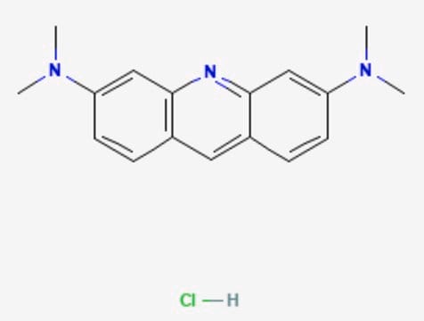
Acridine Orange hemi(zinc chloride) salt

For nucleic acid staining in cells or gels

Synonym(s):

3,6-Bis(dimethylamino)acridine hydrochloride zinc chloride double salt, 3,6-Bis(dimethylamino)acridinium chloride hemi(zinc chloride salt), Basic Orange 14

Sign Into View Organizational & Contract Pricing


About This Item

Linear Formula:
C17H20ClN3 · HCl · 1/2ZnCl2
CAS Number:
Molecular Weight:
369.96
Colour Index Number:
46005
Beilstein:
3734978
EC Number:
MDL number:
UNSPSC Code:
12171500
PubChem Substance ID:
NACRES:
NA.52

Quality Level

product line

BioReagent

form

powder

composition

Dye content, ~80%

technique(s)

nucleic acid detection: suitable

solubility

ethanol: 2 mg/mL
4 mg/mL (2-methoxyethanol (EGME))
water: 6 mg/mL (Forms a clear, dark orange or amber solution at 1mg/mL.)

suitability

suitable for flow cytometry
suitable for microscopy

storage temp.

room temp

SMILES string

Cl[H].Cl[H].Cl[Zn]Cl.CN(C)c1ccc2cc3ccc(cc3nc2c1)N(C)C.CN(C)c4ccc5cc6ccc(cc6nc5c4)N(C)C

InChI

1S/2C17H19N3.4ClH.Zn/c2*1-19(2)14-7-5-12-9-13-6-8-15(20(3)4)11-17(13)18-16(12)10-14;;;;;/h2*5-11H,1-4H3;4*1H;/q;;;;;;+2/p-2

InChI key

RAHGLSRJKRXOSY-UHFFFAOYSA-L

Looking for similar products? Visit Product Comparison Guide

General description

Acridine orange is a metachromatic fluorescent cationic dye that permeates the cell membrane and intercalates DNA and RNA. It allows for visual detection of nucleic acids on agarose and polyacrylamide gels.

Application

Acridine Orange, a cell-permeable metachromatic fluorescent cationic dye that intercalates DNA and RNA, is used in fluorescence and epiflouresence microscopy. Acridine Orange dye has been used to analyze mitochondria and lysosomal content by flow cytometry, characterize multidrug resistance, and measure changes in mitochondrial mass during apoptosis in rat thymocytes.
Suitable for
  • detection of nucleic acids separated by gel electrophoresis
  • fluorescence and epifluorescence microscopy
  • analysis of mitochondria and lysosomes by flow cytometry
  • DNA staining in apoptosis studies

Features and Benefits

  • 120 microM of acridine orange detects 25-50 ng of purified DNA per band in gels
  • differential staining of single- and double-stranded polynucleotides

Principle

Acridine orange intercalates into the nucleic acids of double helix and is detectable as green fluorescence at 530 nm. It binds electrostatically to the phosphate groups in single stranded nucleic acids and is detectable at red fluorescence at 640 nm.

related product

Pictograms

Health hazard

Signal Word

Warning

Hazard Statements

Hazard Classifications

Muta. 2

Storage Class Code

11 - Combustible Solids

WGK

WGK 3

Flash Point(F)

Not applicable

Flash Point(C)

Not applicable

Personal Protective Equipment

dust mask type N95 (US), Eyeshields, Gloves

Certificates of Analysis (COA)

Search for Certificates of Analysis (COA) by entering the products Lot/Batch Number. Lot and Batch Numbers can be found on a product’s label following the words ‘Lot’ or ‘Batch’.

Already Own This Product?

Find documentation for the products that you have recently purchased in the Document Library.

Visit the Document Library

G K McMaster et al.
Proceedings of the National Academy of Sciences of the United States of America, 74(11), 4835-4838 (1977-11-01)
We have developed a simple and rapid system for the denaturation of nucleic acids and their subsequent analysis by gel electrophoresis. RNA and DNA are denatured in 1 M glyoxal (ethanedial) and 50% (vol/vol) dimethyl sulfoxide, at 50 degrees. The
H Baisch et al.
Cell proliferation, 32(5), 303-319 (2000-01-05)
Early indicators of apoptosis in mammalian cells are membrane potential breakdown (loss) in mitochondria (MPLM), chromatin condensation, DNA degradation, and phosphatidylserine exposure (PSE) on the outside plasma membrane. One aim of the present study was to determine the kinetics of
Z Darzynkiewicz et al.
Cytometry, 13(8), 795-808 (1992-01-01)
The present review describes several methods to characterize and differentiate between two different mechanisms of cell death, apoptosis and necrosis. Most of these methods were applied to studies of apoptosis triggered in the human leukemic HL-60 cell line by DNA
F Durrieu et al.
Cytometry, 36(2), 140-149 (1999-11-30)
Some forms of chemoresistance in leukemia may start from failure of tumour cells to successfully undergo apoptosis and Bcl-2 may play a role in this defect. Therefore, we evaluated the Bcl-2 content and synthesis in relation with the apoptotic potential
A B Borisov et al.
Tsitologiia, 24(10), 1233-1237 (1982-10-01)
Using supravital fluorescent staining of lysosomes with Euchrysine 3R, the morphology of these organelles was studied in L cells examined from cultures being at different growth phases in the course of cell cycle and after adipocyte conversion of L cells

Articles

Fluorescence lifetime measurement is advantageous over intensity-based measurements. Applications include fluorescence lifetime assays, sensing and FLI.

Fluorescence lifetime measurement is advantageous over intensity-based measurements. Applications include fluorescence lifetime assays, sensing and FLI.

Fluorescence lifetime measurement is advantageous over intensity-based measurements. Applications include fluorescence lifetime assays, sensing and FLI.

Fluorescence lifetime measurement is advantageous over intensity-based measurements. Applications include fluorescence lifetime assays, sensing and FLI.

Our team of scientists has experience in all areas of research including Life Science, Material Science, Chemical Synthesis, Chromatography, Analytical and many others.

Contact Technical Service