Skip to Content
Merck
All Photos(4)

Key Documents

MABT901

Sigma-Aldrich

Anti-Connexin 43 Antibody, C-terminal Antibody, clone P4G9

clone P4G9, from mouse

Synonym(s):

Gap junction alpha-1 protein, CX43, Gap junction 43 kDa heart protein

Sign Into View Organizational & Contract Pricing


About This Item

UNSPSC Code:
12352203
eCl@ss:
32160702
NACRES:
NA.41
clone:
P4G9, monoclonal
application:
ICC
IF
IHC
IP
species reactivity:
mouse, human, rat
technique(s):
immunocytochemistry: suitable
immunofluorescence: suitable
immunohistochemistry: suitable (paraffin)
immunoprecipitation (IP): suitable
citations:
1

biological source

mouse

Quality Level

antibody form

purified immunoglobulin

antibody product type

primary antibodies

clone

P4G9, monoclonal

species reactivity

mouse, human, rat

species reactivity (predicted by homology)

guinea pig (based on 100% sequence homology), canine (based on 100% sequence homology), rabbit (based on 100% sequence homology), hamster (based on 100% sequence homology), porcine (based on 100% sequence homology), bovine (based on 100% sequence homology), rhesus monkey (based on 100% sequence homology)

technique(s)

immunocytochemistry: suitable
immunofluorescence: suitable
immunohistochemistry: suitable (paraffin)
immunoprecipitation (IP): suitable

isotype

IgG2aκ

NCBI accession no.

UniProt accession no.

shipped in

ambient

target post-translational modification

unmodified

Gene Information

bovine ... Gja1(281193)
dog ... Gja1(403418)
guinea pig ... Gja1(100379273)
human ... GJA1(2697)
mouse ... Gja1(14609)
rabbit ... Gja1(100008935)
rat ... Gja1(24392)
rhesus monkey ... Gja1(714344)

General description

Gap junction alpha-1 protein (UniProt: P08050; also known as Connexin-43, CX43, Gap junction 43 kDa heart protein) is encoded by the Gja1 (also known as Cxn-43) gene (Gene ID: 24392) in rat. The gap junction channels are dodecamers of transmembrane proteins of the connexin family. Connexins usually have a short half-life ( 1.5 - 5 h) and this short half-life is a key element of coupling regulation as it allows very dynamic and acute changes in gap junction regulation and gap junctional communication. In cardiomyocytes three types of connexins have been identified: Connexin 40, 43, and 45. Each of these connexins form channels with unique and specific electrophysiological properties. Connexin-43 is a multi-pass membrane protein and is the most ubiquitous connexin with widespread tissue expression pattern. Connexin-43 can be phosphorylated at multiple sites and phosphorylation is shown to regulate the kinetics of its trafficking, assembly, gating and turnover in a cell-cycle-stage specific manner. Phosphorylation at Ser325, Ser328 and Ser330 by casein kinase I is reported to modulate gap junction assembly while phosphorylation of Ser368 by protein kinase C gamma isoform leads to disassembly of gap junction plaques and inhibition of gap junction activity. During myocardial ischemia connexin-43 is shown to become dephosphorylated that allows gap junctions to be opened and convey the propagation of ischemia injury. In bladder smooth muscle cells, connexin-43 displays a circadian cycle with low levels during the sleep phase.

Specificity

Clone P4G9 specifically detects connexin 43 in human and rat heart. It targets an epitopw within 23 amino acids from the C-terminal end.

Immunogen

Epitope: C-terminus
KLH-conjugated linear peptide corresponding to 23 amino acids from the C-terminal region of rat connexin-43.

Application

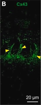
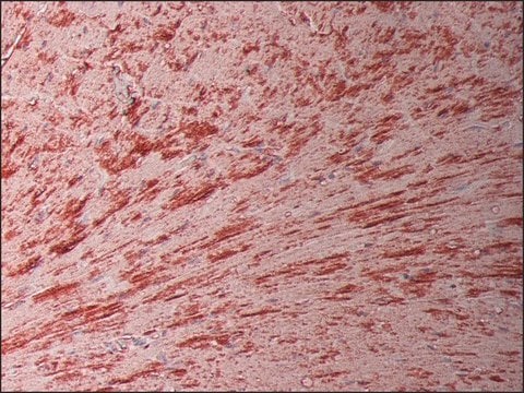
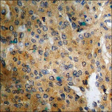
Immunohistochemistry Analysis: A 1:50 dilution from a representative lot detected Connexin 43 in rat heart tissue.

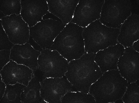
Immunofluorescence Analysis: A representative lot detected Connexin 43 in NRK cells (Courtesy of Joell Solan in Paul Lampe s Lab at the Fred Hutchinson Cancer Research Center, Seattle, WA).

Immunoprecipitation Analysis: A representative lot detected Connexin 43 in Immunoprecipitation applications (Sosinsky, G.E., et. al. (2007). Biochem J. 408(3):375-85).

Immunocytochemistry Analysis: A representative lot detected Connexin 43 in Immunocytochemistry applications (Norris, R.P., et. al. (2008). Development. 135(19):3229-38).

Immunocytochemistry Analysis: A representative lot detected Connexin 43 in Immunocytochemistry applications (Churko, J.M., et. al. (2010). Biochem J. 429(3):473-83).
Research Category
Cell Structure
This mouse monoclonal Anti-Connexin 43, C-terminal, clone P4G9, Cat. No. MABT901 is tested for use in Immunocytochemistry, Immunofluorescence, Immunohistochemistry (Paraffin), and Immunoprecipitation for the detection of Connexin-43.

Quality

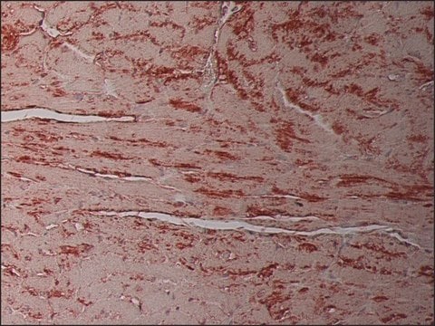
Evaluated by Immunohistochemistry in human heart tissue.

Immunohistochemistry Analysis: A 1:50 dilution of this antibody detected Connexin 43 in human heart tissue.

Target description

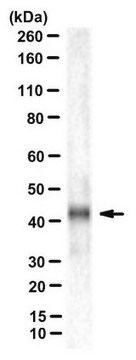
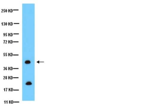
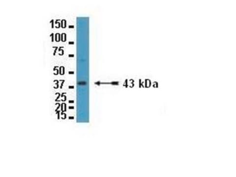
43.03 kDa calculated.

Physical form

Format: Purified
Protein G purified
Purified mouse monoclonal antibody IgG2a in buffer containing 0.1 M Tris-Glycine (pH 7.4), 150 mM NaCl with 0.05% sodium azide.

Storage and Stability

Stable for 1 year at 2-8°C from date of receipt.

Other Notes

Concentration: Please refer to lot specific datasheet.

Disclaimer

Unless otherwise stated in our catalog or other company documentation accompanying the product(s), our products are intended for research use only and are not to be used for any other purpose, which includes but is not limited to, unauthorized commercial uses, in vitro diagnostic uses, ex vivo or in vivo therapeutic uses or any type of consumption or application to humans or animals.

Not finding the right product?  

Try our Product Selector Tool.

Storage Class Code

12 - Non Combustible Liquids

WGK

WGK 1


Certificates of Analysis (COA)

Search for Certificates of Analysis (COA) by entering the products Lot/Batch Number. Lot and Batch Numbers can be found on a product’s label following the words ‘Lot’ or ‘Batch’.

Already Own This Product?

Find documentation for the products that you have recently purchased in the Document Library.

Visit the Document Library

Jessica L Moore et al.
The Journal of cell biology, 222(7) (2023-04-27)
Skin homeostasis is maintained by stem cells, which must communicate to balance their regenerative behaviors. Yet, how adult stem cells signal across regenerative tissue remains unknown due to challenges in studying signaling dynamics in live mice. We combined live imaging

Our team of scientists has experience in all areas of research including Life Science, Material Science, Chemical Synthesis, Chromatography, Analytical and many others.

Contact Technical Service