Skip to Content
Merck
All Photos(3)

Key Documents

MABN495

Sigma-Aldrich

Anti-Aldh1L1 Antibody, clone N103/39

clone N103/39, from mouse

Synonym(s):

Cytosolic 10-formyltetrahydrofolate dehydrogenase, 10-FTHFDH, FDH, Aldehyde dehydrogenase family 1 member L1

FBP-CI

Sign Into View Organizational & Contract Pricing


About This Item

UNSPSC Code:
12352203
eCl@ss:
32160702
NACRES:
NA.41
clone:
N103/39, monoclonal
application:
IF
IHC
WB
species reactivity:
human, rat, mouse
technique(s):
immunofluorescence: suitable
immunohistochemistry: suitable
western blot: suitable
citations:
21

biological source

mouse

Quality Level

antibody form

purified immunoglobulin

antibody product type

primary antibodies

clone

N103/39, monoclonal

species reactivity

human, rat, mouse

technique(s)

immunofluorescence: suitable
immunohistochemistry: suitable
western blot: suitable

isotype

IgG1κ

NCBI accession no.

UniProt accession no.

shipped in

wet ice

target post-translational modification

unmodified

Gene Information

human ... ALDH1L1(10840)

General description

Aldh1L1, also known as 10-formyltetrahydrofolate dehydrogenase (10-FTHFDH), is a multidomain protein that is found in liver cytosol and also serves as a CNS astrocyte marker. Aldh1L1 is comprised of two functionally unrelated domains, an aldehyde dehydrogenase-homologous domain and a folate-binding hydrolase domain. These two domains are thought to be connected by a 100-residue linker. It is known to induce phosphorylation of p53 at Ser6, which is a critical step in stimulating apoptosis.

Specificity

This Aldh1L1 monoclonal antibody does not exhibit cross-reactivity toward Aldh1L2.

Immunogen

Recombinant full-length rat Aldh1L1.

Application

Anti-Aldh1L1, clone N103/39 is an antibody targeting the Aldh1L1 protein, validated for use in Immunofluorescence, Immunohistochemistry, and Western Blotting.
Research Category
Neuroscience
Research Sub Category
Sensory & PNS
Western Blot Analysis: 0.5 µg/mL from a representative lot detected Aldh1L1 in 10 µg of human brain tissue lysate.

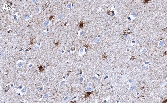
Immunohistochemistry Analysis: A 1:2,000 dilution from a representative lot detected Aldh1L1 rat cerebral cortex tissue and human pons/midbrain tissue.

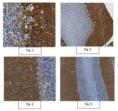
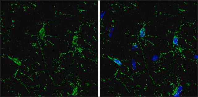
Immunofluorescence Analysis: A representative lot detected Aldh1L1 in rat cortex and cerebellum tissue.

Quality

Evaluated by Western Blot in mouse brain tissue lysate.

Western Blot Analysis: 0.5 µg/mL of this antibody detected Aldh1L1 in 10 µg of mouse brain tissue lysate.

Target description

~99 kDa observed

Physical form

Format: Purified
Protein G Purified
Purified mouse IgG1κ in buffer containing 0.1 M Tris-Glycine (pH 7.4), 150 mM NaCl with 0.05% sodium azide.

Storage and Stability

Stable for 1 year at 2-8°C from date of receipt.

Analysis Note

Control
Mouse brain tissue lysate

Other Notes

Concentration: Please refer to the Certificate of Analysis for the lot-specific concentration.

Disclaimer

Unless otherwise stated in our catalog or other company documentation accompanying the product(s), our products are intended for research use only and are not to be used for any other purpose, which includes but is not limited to, unauthorized commercial uses, in vitro diagnostic uses, ex vivo or in vivo therapeutic uses or any type of consumption or application to humans or animals.

Not finding the right product?  

Try our Product Selector Tool.

Storage Class Code

12 - Non Combustible Liquids

WGK

WGK 1

Flash Point(F)

Not applicable

Flash Point(C)

Not applicable


Certificates of Analysis (COA)

Search for Certificates of Analysis (COA) by entering the products Lot/Batch Number. Lot and Batch Numbers can be found on a product’s label following the words ‘Lot’ or ‘Batch’.

Already Own This Product?

Find documentation for the products that you have recently purchased in the Document Library.

Visit the Document Library

Sheila Espírito-Santo et al.
Glia, 69(6), 1429-1443 (2021-01-27)
Central nervous system (CNS) function depends on precise synaptogenesis, which is shaped by environmental cues and cellular interactions. Astrocytes are outstanding regulators of synapse development and plasticity through contact-dependent signals and through the release of pro- and antisynaptogenic factors. Conversely
New ?- and ?-synuclein immunopathological lesions in human brain.
Surgucheva, I; Newell, KL; Burns, J; Surguchov, A
Acta Neuropathologica Communications null
Gajanan Sathe et al.
Journal of neurochemistry, 156(6), 988-1002 (2020-07-03)
Alzheimer's disease (AD) is a chronic neurodegenerative disease characterized by intracellular formation of neurofibrillary tangles and extracellular deposition of β-amyloid protein (Aβ) in the extracellular matrix. The pathogenesis of AD has not yet been fully elucidated and little is known
Jeremy S Rabinowitz et al.
Proceedings of the National Academy of Sciences of the United States of America, 114(5), E717-E726 (2017-01-18)
Regeneration requires cells to regulate proliferation and patterning according to their spatial position. Positional memory is a property that enables regenerating cells to recall spatial information from the uninjured tissue. Positional memory is hypothesized to rely on gradients of molecules
Maria de Los Angeles Castillo-Rodriguez et al.
Histochemistry and cell biology, 158(1), 15-38 (2022-04-06)
Myelin loss with consecutive axon degeneration and impaired remyelination are the underlying causes of progressive disease in patients with multiple sclerosis. Astrocytes are suggested to play a major role in these processes. The unmasking of distinct astrocyte identities in health

Our team of scientists has experience in all areas of research including Life Science, Material Science, Chemical Synthesis, Chromatography, Analytical and many others.

Contact Technical Service