Skip to Content
Merck
All Photos(4)

Key Documents

AB3786

Sigma-Aldrich

Anti-SFTPC Antibody

CHEMICON®, rabbit polyclonal

Synonym(s):

proSP-C

Sign Into View Organizational & Contract Pricing


About This Item

UNSPSC Code:
12352203
eCl@ss:
32160702
NACRES:
NA.41
clone:
polyclonal
application:
IHC (p)
WB
species reactivity:
mouse, rat
technique(s):
immunohistochemistry (formalin-fixed, paraffin-embedded sections): suitable
western blot: suitable
citations:
367

Product Name

Anti-Prosurfactant Protein C (proSP-C) Antibody, serum, Chemicon®

biological source

rabbit

Quality Level

antibody form

serum

antibody product type

primary antibodies

clone

polyclonal

species reactivity

mouse, rat

manufacturer/tradename

Chemicon®

technique(s)

immunohistochemistry (formalin-fixed, paraffin-embedded sections): suitable
western blot: suitable

NCBI accession no.

UniProt accession no.

shipped in

dry ice

target post-translational modification

unmodified

Gene Information

General description

Pulmonary surfactant is a complex mixture of phospholipids and proteins secreted from airway epithelial cells (type II cells) in alveoli which reduces the surface tension at the alveolar air liquid interface, providing alveolar stability necessary for normal ventilation. Four distinct proteins isolated from pulmonary surfactant are termed surfactant proteins A, B, C and D. SP A (28 - 36 kDa) and SP D (43 kDa) are collagenous carbohydrate binding proteins, whereas SP B (8 - 9 kDa) and SP C (4 kDa) are non collagenous hydrophobic proteins. SP C is a fragment of a much larger precursor protein of 21 kDa. The precursor contains an exceedingly hydrophobic region of 34 amino acids that comprises most of the mature SP C, and a unique poly-valine domain.

Specificity

Expected to cross-react with human.
Reacts strongly with proSP-C from human and mouse. Immunoprecipitates the 21 kDa proprotein as well as processing intermediates of M.W. 26 kDa, 21 kDa, 16 kDa and 14 kDa. Preincubation of the antiserum with the fusion protein results in ablation of immunostaining. On human tissue immunostains alveolar Type II cells in normal lung. The antibody also recognizes the aberrant form of proSP-C (6-12 kDa peptide) found in BAL and in alveolar proteinosis material on sections of lung samples from human patients with hereditary SP-B deficiency. On mouse tissue immunostains alveolar Type II cells in normal lung.

Immunogen

GST-tagged recombinant protein corresponding to the N-terminal region of human Prosurfactant Protein C (proSP-C).

Application

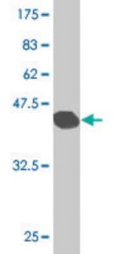
  • Western Blotting Analysis: A 1:500 dilution from a representative lot detected Prosurfactant Protein C (proSP-C) in mouse lung tissue lysate and GST-fusion Prosurfactant Protein C fragment.
  • ELISA Analysis: A 1:50,000 dilution from a representative lot detected GST-fusion Prosurfactant Protein C fragment.
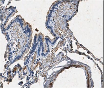
  • Immunohistochemistry (Paraffin) Analysis: A 1:2,000 dilution from a representative lot detected Prosurfactant Protein C (proSP-C) in mouse lung tissue sections.
  • Note: Actual optimal working dilutions must be determined by end user as specimens, and experimental conditions may vary with the end user.
Research Category
Metabolism
Research Sub Category
Ion & Transport Channels
This rabbit polyclonal Anti-Prosurfactant Protein C (proSP-C), Cat. No. AB3786 is tested for use in Western Blotting, Immunohistochemistry (Paraffin) and ELISA, for the detection of Prosurfactant Protein C (proSP-C).

Quality

Evaluated by Western Blotting in rat lung tissue lysate. Western Blotting Analysis: A 1:500 dilution of this antibody detected Prosurfactant Protein C (proSP-C) in rat lung tissue lysate.

Target description

20 kDa observed. 21 kDa calculated. Uncharacterized bands may be observed in some lysate(s).

Linkage

Replaces: 07-647

Physical form

Rabbit polyclonal antibody in serum with 0.05% sodium azide.
Unpurified

Storage and Stability

Stable at -20ºC in undiluted aliquots for up to twelve months after date of receipt. Avoid repeated freeze/thaw cycles.

Analysis Note

Control
Mouse lung epithelial cells.

Other Notes

Concentration: Please refer to the Certificate of Analysis for the lot-specific concentration.

Legal Information

CHEMICON is a registered trademark of Merck KGaA, Darmstadt, Germany

Disclaimer

Unless otherwise stated in our catalog or other company documentation accompanying the product(s), our products are intended for research use only and are not to be used for any other purpose, which includes but is not limited to, unauthorized commercial uses, in vitro diagnostic uses, ex vivo or in vivo therapeutic uses or any type of consumption or application to humans or animals.

Not finding the right product?  

Try our Product Selector Tool.

Storage Class Code

12 - Non Combustible Liquids

WGK

WGK 1

Flash Point(F)

Not applicable

Flash Point(C)

Not applicable


Certificates of Analysis (COA)

Search for Certificates of Analysis (COA) by entering the products Lot/Batch Number. Lot and Batch Numbers can be found on a product’s label following the words ‘Lot’ or ‘Batch’.

Already Own This Product?

Find documentation for the products that you have recently purchased in the Document Library.

Visit the Document Library

Evidence for type II cells as cells of origin of K-Ras-induced distal lung adenocarcinoma.
Xu, X; Rock, JR; Lu, Y; Futtner, C; Schwab, B; Guinney, J; Hogan, BL; Onaitis, MW
Proceedings of the National Academy of Sciences of the USA null
EGF receptor signaling is essential for k-ras oncogene-driven pancreatic ductal adenocarcinoma.
Navas, C; Hernandez-Porras, I; Schuhmacher, AJ; Sibilia, M; Guerra, C; Barbacid, M
Cancer Cell null
Christian C Hofer et al.
American journal of physiology. Lung cellular and molecular physiology, 308(7), L628-L638 (2015-01-18)
Influenza viruses cause acute respiratory disease of great importance to public health. Alveolar type II (ATII) respiratory epithelial cells are central to normal lung function and are a site of influenza A virus replication in the distal lung. However, the
Julia A Paxson et al.
PloS one, 6(8), e23232-e23232 (2011-09-14)
While aging leads to a reduction in the capacity for regeneration after pneumonectomy (PNX) in most mammals, this biological phenomenon has not been characterized over the lifetime of mice. We measured the age-specific (3, 9, 24 month) effects of PNX
Oliver Delgado et al.
PloS one, 6(7), e22023-e22023 (2011-07-16)
While the adult murine lung utilizes multiple compartmentally restricted progenitor cells during homeostasis and repair, much less is known about the progenitor cells from the human lung. Translating the murine stem cell model to humans is hindered by anatomical differences

Our team of scientists has experience in all areas of research including Life Science, Material Science, Chemical Synthesis, Chromatography, Analytical and many others.

Contact Technical Service