Skip to Content
Merck
All Photos(1)

Documents

C5176

Sigma-Aldrich

Monoclonal Anti-Cytokeratin Peptide 4 antibody produced in mouse

clone 6B10, ascites fluid

Synonym(s):

Anti-CK-4, Anti-CK4, Anti-CYK4, Anti-K4, Anti-WSN1

Sign Into View Organizational & Contract Pricing


About This Item

MDL number:
UNSPSC Code:
12352203
NACRES:
NA.41

biological source

mouse

Quality Level

conjugate

unconjugated

antibody form

ascites fluid

antibody product type

primary antibodies

clone

6B10, monoclonal

mol wt

antigen 59 kDa

contains

15 mM sodium azide

species reactivity

rabbit, feline, hamster, goat, guinea pig, human, canine, sheep

technique(s)

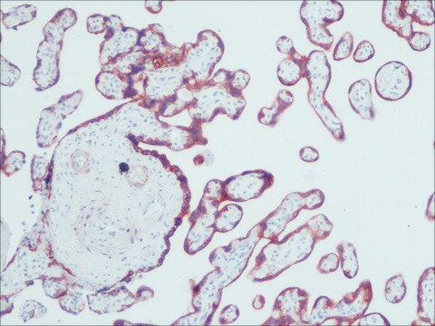
immunohistochemistry (formalin-fixed, paraffin-embedded sections): suitable
immunohistochemistry (frozen sections): suitable
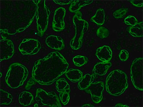
indirect immunofluorescence: 1:300 using formalin-fixed, paraffin-embedded sections of human or animal tissue
microarray: suitable
western blot: suitable

isotype

IgG1

UniProt accession no.

shipped in

dry ice

storage temp.

−20°C

target post-translational modification

unmodified

Gene Information

human ... KRT4(3851)

General description

Cytokeratins are intermediary filaments that are expressed during epithelial development. Cytokeratin-4 is largely found in non-epidermal, stratified epithelial tissues such as larynx, pharynx, vagina, exocervix and epiglottis among several others . Cytokeratin-4 has been implicated in Streptococcus agalactiae infection and genetic mutations in Cytokeratin-4 have been linked to white sponge naevus . Monoclonal Anti-Cytokeratin Peptide 4 antibody is specific for cytokeratin 4 in goats, hamsters, guinea pigs, cats, sheep, humans, rabbits, and dogs. The antibody weakly stains ciliated pseudostratified epithelium of bronchi. Ducts of skin sweat glands and prostate are focally stained by the antibody. Some endocervical columnar cells may also react with the antibody. This antibody is reactive with methanol or acetone fixed frozen sections, and with protease-digested formalin fixed paraffin embedded human tissues. Embedded methacarn fixed material can also be used for cytokeratin 4 demonstration.

Specificity

The antibody reacts with non-cornifying squamous epithelium (e.g., exocervix, esophagus and cornea). Ciliated pseudostratified epithelium of bronchi is weakly stained. Ducts of sweat glands and prostate are focally stained. The antibody may react with some endocervical columnar cells.

Immunogen

cytokeratin from human esophagus.

Application

Monoclonal Anti-Cytokeratin Peptide 4 antibody is suitable for use in a broad range of immunoassays such as immunohistochemistry (frozen or formalin-fixed, paraffin-embedded sections), immunoblot, dot blot, and indirect immunofluorescence (1:300 using formalin-fixed, paraffin-embedded sections of human or animal tissue). The antibody may also be used for western blot and microarray.

Disclaimer

Unless otherwise stated in our catalog or other company documentation accompanying the product(s), our products are intended for research use only and are not to be used for any other purpose, which includes but is not limited to, unauthorized commercial uses, in vitro diagnostic uses, ex vivo or in vivo therapeutic uses or any type of consumption or application to humans or animals.

Not finding the right product?  

Try our Product Selector Tool.

Storage Class Code

10 - Combustible liquids

WGK

nwg

Flash Point(F)

Not applicable

Flash Point(C)

Not applicable


Certificates of Analysis (COA)

Search for Certificates of Analysis (COA) by entering the products Lot/Batch Number. Lot and Batch Numbers can be found on a product’s label following the words ‘Lot’ or ‘Batch’.

Already Own This Product?

Find documentation for the products that you have recently purchased in the Document Library.

Visit the Document Library

Dianlei Guo et al.
Experimental eye research, 180, 208-225 (2018-12-28)
Ocular surface disease is one major type of eye diseases. Different etiologies trigger distinct pathological responses of the ocular surface. We previously reported that genetically engineered mice with ablation of Prickle 1 manifested precocious eyelid opening with ensuing cornea dysplasia.
Tamsin R Sheen et al.
Journal of bacteriology, 193(24), 6834-6842 (2011-10-11)
Streptococcus agalactiae (group B streptococcus [GBS]) is a Gram-positive bacterium found in the female rectovaginal tract and is capable of producing severe disease in susceptible hosts, including newborns and pregnant women. The vaginal tract is considered a major reservoir for
S-C Chao et al.
The British journal of dermatology, 148(6), 1125-1128 (2003-06-28)
White sponge naevus (WSN) is a rare, autosomal dominant disorder that predominantly affects noncornified stratified squamous epithelia, most commonly the buccal mucosa. Clinically, WSN manifests as thickened spongy mucosa with a white opalescent tint in the mouth and may be
Ulrike Samen et al.
Infection and immunity, 75(11), 5405-5414 (2007-08-22)
Streptococcus agalactiae is frequently the cause of bacterial sepsis and meningitis in neonates. In addition, it is a commensal bacterium that colonizes the mammalian gastrointestinal tract. During its commensal and pathogenic lifestyles, S. agalactiae colonizes and invades a number of
R E Leube et al.
The Journal of cell biology, 106(4), 1249-1261 (1988-04-01)
A number of human cytokeratins are expressed during the development of stratified epithelia from one-layered polar epithelia and continue to be expressed in several adult epithelial tissues. For studies of the regulation of the synthesis of stratification-related cytokeratins in internal

Our team of scientists has experience in all areas of research including Life Science, Material Science, Chemical Synthesis, Chromatography, Analytical and many others.

Contact Technical Service