Skip to Content
Merck
All Photos(2)

Key Documents

07-1213

Sigma-Aldrich

Anti-IP3 Receptor 1 Antibody

serum, from rabbit

Synonym(s):

IP3 receptor isoform 1, Type 1 InsP3 receptor, Type 1 inositol 1,4,5-trisphosphate receptor, inositol 1,4,5-triphosphate receptor, type 1, spinocerebellar ataxia 15, spinocerebellar ataxia 16

Sign Into View Organizational & Contract Pricing


About This Item

UNSPSC Code:
12352203
eCl@ss:
32160702
NACRES:
NA.41
clone:
polyclonal
application:
IHC
WB
species reactivity:
mouse, human
technique(s):
immunohistochemistry: suitable (paraffin)
western blot: suitable
citations:
5

biological source

rabbit

Quality Level

antibody form

serum

antibody product type

primary antibodies

clone

polyclonal

species reactivity

mouse, human

species reactivity (predicted by homology)

canine, rat

technique(s)

immunohistochemistry: suitable (paraffin)
western blot: suitable

isotype

IgG

UniProt accession no.

shipped in

wet ice

target post-translational modification

unmodified

Gene Information

dog ... Itpr1(476548)
human ... ITPR1(3708)
mouse ... Itpr1(16438)
rat ... Itpr1(25262)

General description

IP3 Receptor 1 is an intracellular channel that when stimulated by inositol 1,4,5-triphosphate mediates the release of calcium from the endoplasmic reticulum. Defects within the gene ITPR1 cause a group of cerebellar disorders known as spinocerebellar ataxia type 15 (SCA15). This receptor is widely expressed in tissue.

Specificity

The antibody reacts with the 300 kDa IP3 Receptor 1 protein.

Immunogen

Synthetic peptide from the N-terminal of human IP3 Receptor 1.

Application

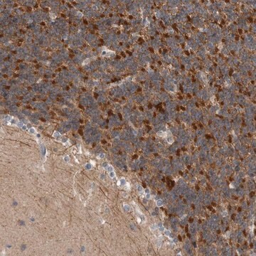
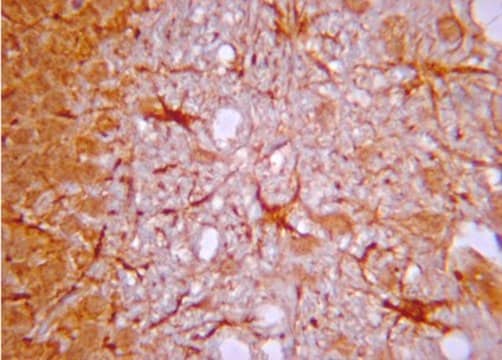
Immunohistochemistry Analysis: A 1:100 dilution of this antibody detected IP3 Receptor 1 in mouse cerebellum tissue.
Research Category
Signaling
Research Sub Category
Lipid Signaling
This Anti-IP3 Receptor 1 Antibody is validated for use in IH(P), WB for the detection of IP3 Receptor 1.

Quality

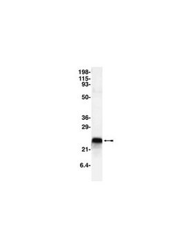
Evaluated by Western Blot in C2C12 cell lysates.
Western Blot Analysis: 1:500 dilution of this lot detected IP3 RECPT 1 on 10 ug of C2C12 lysates.

Target description

Approx. 300 kDa

Linkage

Replaces: AB9072

Physical form

Rabbit serum. Liquid
Unpurified rabbit serum

Storage and Stability

Maintain at -20°C in undiluted aliquots for up to 1 year date of receipt. Avoid repeated freeze/thaw cycles.

Analysis Note

Control
C2C12 cell lysates

Other Notes

Concentration: Please refer to the Certificate of Analysis for the lot-specific concentration.

Disclaimer

Unless otherwise stated in our catalog or other company documentation accompanying the product(s), our products are intended for research use only and are not to be used for any other purpose, which includes but is not limited to, unauthorized commercial uses, in vitro diagnostic uses, ex vivo or in vivo therapeutic uses or any type of consumption or application to humans or animals.

Not finding the right product?  

Try our Product Selector Tool.

Storage Class Code

10 - Combustible liquids

WGK

WGK 1

Flash Point(F)

Not applicable

Flash Point(C)

Not applicable


Certificates of Analysis (COA)

Search for Certificates of Analysis (COA) by entering the products Lot/Batch Number. Lot and Batch Numbers can be found on a product’s label following the words ‘Lot’ or ‘Batch’.

Already Own This Product?

Find documentation for the products that you have recently purchased in the Document Library.

Visit the Document Library

Vera Khodzhaeva et al.
Frontiers in immunology, 12, 723683-723683 (2021-08-31)
Mitofusin 2 (MFN2) is a mitochondrial outer membrane GTPase, which modulates mitochondrial fusion and affects the interaction between endoplasmic reticulum and mitochondria. Here, we explored how MFN2 influences mitochondrial functions and inflammatory responses towards zymosan in primary human macrophages. A
Michael R Butler et al.
The Journal of biological chemistry, 292(27), 11189-11205 (2017-05-13)
Endoplasmic reticulum (ER) stress and mislocalization of improperly folded proteins have been shown to contribute to photoreceptor death in models of inherited retinal degenerative diseases. In particular, mice with cone cyclic nucleotide-gated (CNG) channel deficiency, a model for achromatopsia, display
Xiang Liu et al.
Molecular medicine reports, 14(6), 5435-5442 (2016-11-15)
Isoflurane is a commonly used inhaled anesthetic, which induces apoptosis of SH‑SY5Y cells in a dose‑ and time‑dependent manner; however, the underlying mechanisms remain unknown. The authors of the present study hypothesized that a mutation in β‑amyloid precursor protein (APP)
Leonora Szabo et al.
EMBO reports, 24(8), e57499-e57499 (2023-07-04)
Abnormal tau protein impairs mitochondrial function, including transport, dynamics, and bioenergetics. Mitochondria interact with the endoplasmic reticulum (ER) via mitochondria-associated ER membranes (MAMs), which coordinate and modulate many cellular functions, including mitochondrial cholesterol metabolism. Here, we show that abnormal tau
Manon Moulis et al.
Cells, 8(6) (2019-06-28)
Mitochondria-associated ER membranes (MAMs) are crucial for lipid transport and synthesis, calcium exchange, and mitochondrial functions, and they also act as signaling platforms. These contact sites also play a critical role in the decision between autophagy and apoptosis with far

Our team of scientists has experience in all areas of research including Life Science, Material Science, Chemical Synthesis, Chromatography, Analytical and many others.

Contact Technical Service