Skip to Content
Merck
All Photos(2)

Documents

A4187

Sigma-Aldrich

Anti-Goat IgG (whole molecule)–Alkaline Phosphatase antibody produced in rabbit

affinity isolated antibody, buffered aqueous glycerol solution

Synonym(s):

Rabbit Anti-Goat IgG (whole molecule)–AP

Sign Into View Organizational & Contract Pricing


About This Item

MDL number:
UNSPSC Code:
12352203
NACRES:
NA.46

biological source

rabbit

conjugate

alkaline phosphatase conjugate

antibody form

affinity isolated antibody

antibody product type

secondary antibodies

clone

polyclonal

form

buffered aqueous glycerol solution

species reactivity

goat

technique(s)

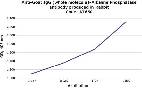
direct ELISA: 1:30,000
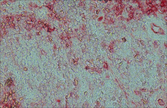
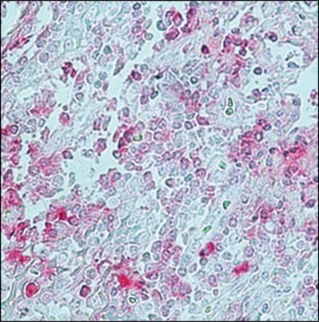
immunohistochemistry (formalin-fixed, paraffin-embedded sections): 1:50
western blot: 1:30,000

shipped in

wet ice

storage temp.

2-8°C

target post-translational modification

unmodified

Looking for similar products? Visit Product Comparison Guide

Related Categories

General description

IgG antibodies regulate several functions such as complement activation and phagocytosis. Thus they play a crucial role in facilitating cytological immune responses. Polyclonal anti-goat IgG (whole molecule)–alkaline phosphatase antibody (diluted 1:15,000) can be used as a secondary antibody for immunoblotting of Sall4. This antibody can also be used in immunohistochemistry (diluted1:400) . Rabbit anti-goat IgG antibody reacts specifically with goat IgG and normal goat serum.
Primary goat antibodies are often used to study target proteins for various clinical and research purposes. Thus, secondary anti-goat antibody conjugated to a detectable substrate can be used to facilitate the accurate detection and localization of target proteins.

Specificity

Binds all goat Igs

Immunogen

Purified goat IgG.

Application

Anti-Goat IgG (whole molecule)-Alkaline Phosphatase antibody is suitable for use in immunoblotting. The product can also be used for direct ELISA (1:30,000) and immunohistochemistry (1:50 using formalin-fixed, paraffin-embedded sections).
Detection of TLR2 and TLR4 in protein extracts from epidermal keratinocytes was performed by western blot using alkaline phosphatase conjugated rabbit anti-goat IgG as the secondary at a 1:2500 dilution.

Physical form

Solution in 0.05 M Tris buffer, pH 8.0, containing 1 mM MgCl2, 1% bovine serum albumin, 50% glycerol and 15 mM sodium azide.

Disclaimer

Unless otherwise stated in our catalog or other company documentation accompanying the product(s), our products are intended for research use only and are not to be used for any other purpose, which includes but is not limited to, unauthorized commercial uses, in vitro diagnostic uses, ex vivo or in vivo therapeutic uses or any type of consumption or application to humans or animals.

Not finding the right product?  

Try our Product Selector Tool.

Storage Class Code

10 - Combustible liquids

WGK

WGK 2


Certificates of Analysis (COA)

Search for Certificates of Analysis (COA) by entering the products Lot/Batch Number. Lot and Batch Numbers can be found on a product’s label following the words ‘Lot’ or ‘Batch’.

Already Own This Product?

Find documentation for the products that you have recently purchased in the Document Library.

Visit the Document Library

Andor Pivarcsi et al.
International immunology, 15(6), 721-730 (2003-05-17)
Keratinocytes have the ability to kill pathogenic fungi and bacteria by producing antimicrobial substances. Recent studies suggest that microbial components use signaling molecules of the human Toll-like receptor (TLR) family to transduce signals in various cells. Here we provide evidence
Menhaj et al.
Planta, 209(4), 406-413 (1999-11-07)
An antibody was raised against the protein HL#2 which is a nuclear-encoded light-stress-induced protein of barley (Hordeum vulgare L.). The expression of the mRNA and the protein of HL#2 was determined under the influence of high light and methyl jasmonate.
Minoru Omi et al.
The Journal of comparative neurology, 519(13), 2615-2621 (2011-04-15)
The optic tectum is a visual center of nonmammalian vertebrates that receives retinal fibers in a retinotopic manner. It has been accepted that retinal fibers project to some superficial laminae of the tectum, but do not go through lamina g
L Moysset et al.
Planta, 213(4), 565-574 (2001-09-15)
The intracellular localization of phytochrome in the pulvini of Robinia pseudoacacia L. was analyzed by immunogold electron microscopy after red (R; 15 min) and far-red (FR; 5 min) irradiation 2 h after the beginning of the photoperiod. Screening of the
Raquel Alvarado et al.
FASEB journal : official publication of the Federation of American Societies for Experimental Biology, 31(1), 85-95 (2016-09-30)
The NLRP3 inflammasome is a multimeric protein complex that controls the production of IL-1β, a cytokine that influences the development of both innate and adaptive immune responses. Helminth parasites secrete molecules that interact with innate immune cells, modulating their activity

Our team of scientists has experience in all areas of research including Life Science, Material Science, Chemical Synthesis, Chromatography, Analytical and many others.

Contact Technical Service