MABT397
Anti-CEACAM1/CD66a Antibody, clone B3-17
clone B3-17, from mouse
Synonym(s):
Carcinoembryonic antigen-related cell adhesion molecule 1, Biliary glycoprotein 1, BGP-1, CD66a
About This Item
FACS
ICC
IHC
WB
flow cytometry: suitable
immunocytochemistry: suitable
immunohistochemistry: suitable (paraffin)
western blot: suitable
Recommended Products
biological source
mouse
Quality Level
antibody form
purified immunoglobulin
antibody product type
primary antibodies
clone
B3-17, monoclonal
species reactivity
human
technique(s)
ELISA: suitable
flow cytometry: suitable
immunocytochemistry: suitable
immunohistochemistry: suitable (paraffin)
western blot: suitable
isotype
IgG1κ
NCBI accession no.
UniProt accession no.
shipped in
wet ice
target post-translational modification
unmodified
Gene Information
human ... CEACAM1(634)
Related Categories
General description
Immunogen
Application
Flow Cytometry Analysis: 10 µg/mL from a representative lot detected the exogenously expressed human CEACAM1/CD66a on the surface of transfected HeLa cells (Courtesy of Dr. B. Singer, University Duisburg-Essen, Germany).
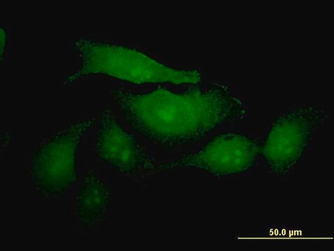
Immunocytochemistry Analysis: 10 µg/mL from a representative lot detected CEACAM1/CD66a on the surface of HT-29 human colorectal carcinoma cells (Courtesy of Dr. B. Singer, University Duisburg-Essen, Germany).
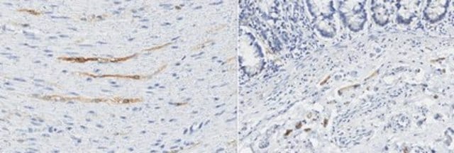
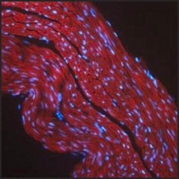
Immunohistochemistry Analysis: 10 µg/mL from a representative lot detected CEACAM1/CD66a immunoreactivity in human jejunum tissue (Courtesy of Dr. B. Singer, University Duisburg-Essen, Germany).
Western Blotting Analysis: 10 µg/mL from a representative lot detected the exogensously expressed human CEACAM1/CD66a extracellular domain-Fc fusion in lysates from transfected cells (Courtesy of Dr. B. Singer, University Duisburg-Essen, Germany).
Flow Cytometry Analysis: Representative lots, either unconjugated or FITC-conjugated, detected CECAM1 immunoreactivity on the surface of human peripheral blood naïve and memory B-cells (Khairnar, V., et al. (2015). Nat. Commun. 6:6217; Seifert, M., et al. (2015). Proc. Natl. Acad. Sci. 112(6):E546-555).
Flow Cytometry Analysis: A representative lot detected CEACAM1/CD66a induction on the surface of normal human bronchial epithelial (NHBE) cells stimulated with poly(I:C) or interferons (Klaile, E., et al. (2013) Respir Res. 14:85).
Flow Cytometry Analysis: A representative lot detected CEACAM1/CD66a expression on the surface of 2 day-starved human epithelial (HT29, T102/3) and endothelial (AS-M.5) cells, as well as multivesicular bodies (MVBs) derived from these cells (Muturi H.T., et al. (2013) PLoS One. 8(9):e74654).
Immunocytochemistry Analysis: A representative lot detected CEACAM1/CD66a immunoreactivity on the surface of normal human bronchial epithelial (NHBE) cells by indirect immunofluorescence staining (Klaile, E., et al. (2013) Respir Res. 14:85).
Immunohistochemistry Analysis: A representative lot detected CEACAM1/CD66a in paraffin-embedded human lung cancer tissue sections (Klaile, E., et al. (2013) Respir Res. 14:85).
Western Blot Analysis: A representative lot detected CEACAM1/CD66a in 2 day-starved AS-M.5 human endothelialn cells and AS-M.5-derived multivesicular bodies (MVBs) (Muturi H.T., et al. (2013) PLoS One. 8(9):e74654).
Quality
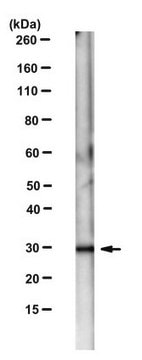
Western Blotting Analysis: 2 µg/mL of this antibody detected CEACAM1/CD66a in 10 µg of HepG2 cell lysate.
Target description
Physical form
Other Notes
Not finding the right product?
Try our Product Selector Tool.
Storage Class Code
12 - Non Combustible Liquids
WGK
WGK 1
Flash Point(F)
Not applicable
Flash Point(C)
Not applicable
Certificates of Analysis (COA)
Search for Certificates of Analysis (COA) by entering the products Lot/Batch Number. Lot and Batch Numbers can be found on a product’s label following the words ‘Lot’ or ‘Batch’.
Already Own This Product?
Find documentation for the products that you have recently purchased in the Document Library.
Our team of scientists has experience in all areas of research including Life Science, Material Science, Chemical Synthesis, Chromatography, Analytical and many others.
Contact Technical Service