Skip to Content
Merck
All Photos(6)

Key Documents

HPA018113

Sigma-Aldrich

Anti-TTBK2 antibody produced in rabbit

enhanced validation

Prestige Antibodies® Powered by Atlas Antibodies, affinity isolated antibody, buffered aqueous glycerol solution

Synonym(s):

Anti-Tau-tubulin kinase 2

Sign Into View Organizational & Contract Pricing


About This Item

UNSPSC Code:
12352203
Human Protein Atlas Number:
NACRES:
NA.43

biological source

rabbit

Quality Level

conjugate

unconjugated

antibody form

affinity isolated antibody

antibody product type

primary antibodies

clone

polyclonal

product line

Prestige Antibodies® Powered by Atlas Antibodies

form

buffered aqueous glycerol solution

species reactivity

human

enhanced validation

recombinant expression
Learn more about Antibody Enhanced Validation

technique(s)

immunohistochemistry: 1:500-1:1000
western blot: 0.04-0.4 μg/mL

immunogen sequence

TTEPLDVTKTQTFSVVPNQDKNNEIMKLLTVGTSEISSRDIDPHVEGQIGQVAEMQKNKISKDDDIMSEDLPGHQGDLSTFLHQEGKREKITPRNGELFHCVS

UniProt accession no.

shipped in

wet ice

storage temp.

−20°C

target post-translational modification

unmodified

Gene Information

human ... TTBK2(146057)

General description

Tau-tubulin kinase 2 (TTBK2) is a serine/threonine kinase expressed in various tissues and it belongs to the casein kinase 1 family. The gene encoding it is localized on human chromosome 15q15.2.

Immunogen

Tau-tubulin kinase 2 recombinant protein epitope signature tag (PrEST)

Application

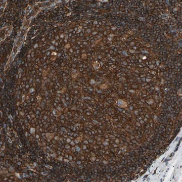
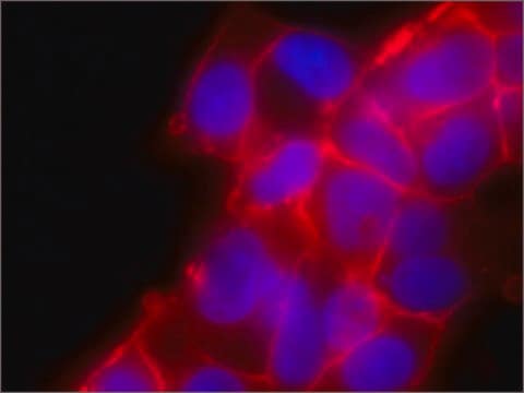
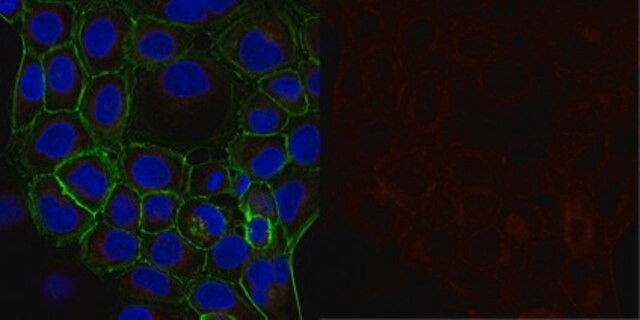
Anti-TTBK2 antibody produced in rabbit, a Prestige Antibody, is developed and validated by the Human Protein Atlas (HPA) project . Each antibody is tested by immunohistochemistry against hundreds of normal and disease tissues. These images can be viewed on the Human Protein Atlas (HPA) site by clicking on the Image Gallery link. The antibodies are also tested using immunofluorescence and western blotting. To view these protocols and other useful information about Prestige Antibodies and the HPA, visit sigma.com/prestige. Anti-TTBK2 antibody produced in rabbit has been used for immunoprecipitation.
These images and the collection of this vast data set can be viewed on the Human Protein Atlas (HPA) site by clicking on the Image Gallery link. We also provide Prestige Antibodies® protocols and other useful information.

Biochem/physiol Actions

Tau-tubulin kinase 2 (TTBK2) forms a complex with centrosomal protein 164kDa (Cep164) of the distal appendages. It also stimulates processes involved in ciliogenesis and recruitment of intraflagellar transport proteins. It has been shown that TTBK2 works upstream of the Cep164 gene and functions in the assemblage of distal appendages. The kinase also regulates the glucose carrier sodium-glucose linked transporter 1 (SGLT1) and increases the number of SGLT1 proteins in the cell membrane. Mutations in the gene encoding it is linked to spinocerebellar ataxia type 11.

Features and Benefits

Prestige Antibodies® are highly characterized and extensively validated antibodies with the added benefit of all available characterization data for each target being accessible via the Human Protein Atlas portal linked just below the product name at the top of this page. The uniqueness and low cross-reactivity of the Prestige Antibodies® to other proteins are due to a thorough selection of antigen regions, affinity purification, and stringent selection. Prestige antigen controls are available for every corresponding Prestige Antibody and can be found in the linkage section.

Every Prestige Antibody is tested in the following ways:
  • IHC tissue array of 44 normal human tissues and 20 of the most common cancer type tissues.
  • Protein array of 364 human recombinant protein fragments.

Linkage

Corresponding Antigen APREST73473

Physical form

Solution in phosphate-buffered saline, pH 7.2, containing 40% glycerol and 0.02% sodium azide

Legal Information

Prestige Antibodies is a registered trademark of Merck KGaA, Darmstadt, Germany

Disclaimer

Unless otherwise stated in our catalog or other company documentation accompanying the product(s), our products are intended for research use only and are not to be used for any other purpose, which includes but is not limited to, unauthorized commercial uses, in vitro diagnostic uses, ex vivo or in vivo therapeutic uses or any type of consumption or application to humans or animals.

Not finding the right product?  

Try our Product Selector Tool.

Storage Class Code

10 - Combustible liquids

WGK

WGK 1

Flash Point(F)

Not applicable

Flash Point(C)

Not applicable

Personal Protective Equipment

dust mask type N95 (US), Eyeshields, Gloves

Choose from one of the most recent versions:

Certificates of Analysis (COA)

Lot/Batch Number

Don't see the Right Version?

If you require a particular version, you can look up a specific certificate by the Lot or Batch number.

Already Own This Product?

Find documentation for the products that you have recently purchased in the Document Library.

Visit the Document Library

Tetsuo Kobayashi et al.
Frontiers in cell and developmental biology, 8, 587691-587691 (2020-12-01)
Primary cilia are hair-like projections that protrude from most mammalian cells and mediate various extracellular signaling pathways. Pancreatic ductal adenocarcinoma (PDAC) cells are known to lose their primary cilia, but the relevance of this phenomenon remains unclear. In this study
Margaret P Adam et al.
GeneReviews(?), 2008 22 (Updated 20 (2013-03-07)
Spinocerebellar ataxia type 11 (SCA11) is characterized by progressive cerebellar ataxia and abnormal eye signs (jerky pursuit, horizontal and vertical nystagmus). Pyramidal features, peripheral neuropathy, and dystonia are seen on occasion. Four families have been reported to date: one each
Ondrej Bernatik et al.
Frontiers in cell and developmental biology, 9, 623753-623753 (2021-03-16)
Primary cilia act as crucial regulators of embryo development and tissue homeostasis. They are instrumental for modulation of several signaling pathways, including Hedgehog, WNT, and TGF-β. However, gaps exist in our understanding of how cilia formation and function is regulated.
Yuriko Sobu et al.
Proceedings of the National Academy of Sciences of the United States of America, 118(10) (2021-03-04)
Mutations that activate LRRK2 protein kinase cause Parkinson's disease. We showed previously that Rab10 phosphorylation by LRRK2 enhances its binding to RILPL1, and together, these proteins block cilia formation in a variety of cell types, including patient derived iPS cells.
Ioana Alesutan et al.
Cellular physiology and biochemistry : international journal of experimental cellular physiology, biochemistry, and pharmacology, 30(2), 458-465 (2012-07-21)
The Tau-tubulin-kinase 2 (TTBK2) is a serine/threonine kinase expressed in various tissues including tumors. Up-regulation of TTBK2 increases resistance of tumor cells against antiangiogenic treatment and confers cell survival. Tumor cell survival critically depends on cellular uptake of glucose, which

Our team of scientists has experience in all areas of research including Life Science, Material Science, Chemical Synthesis, Chromatography, Analytical and many others.

Contact Technical Service