Skip to Content
Merck
All Photos(1)

Key Documents

ABN1686

Sigma-Aldrich

Anti-Rootletin

from chicken, purified by affinity chromatography

Synonym(s):

Ciliary rootlet coiled-coil protein

Sign Into View Organizational & Contract Pricing


About This Item

UNSPSC Code:
12352203
eCl@ss:
32160702
NACRES:
NA.41

biological source

chicken

Quality Level

antibody form

affinity isolated antibody

antibody product type

primary antibodies

clone

polyclonal

purified by

affinity chromatography

species reactivity

human, porcine, mouse

technique(s)

western blot: suitable

isotype

IgY

NCBI accession no.

UniProt accession no.

shipped in

ambient

target post-translational modification

unmodified

Gene Information

mouse ... Crocc(230872)

General description

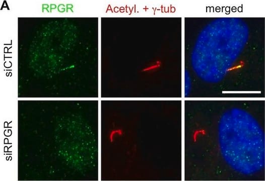
Rootletin (UniProt: Q8CJ40; also known as Ciliary rootlet coiled-coil protein) is encoded by the Crocc (also known as Kiaa0445) gene (Gene ID: 230872) in murine species. Rootletin is a major structural component of the ciliary rootlet, a cytoskeletal-like structure in ciliated cells that originates from the basal body at the proximal end of a cilium and extends proximally toward the cell nucleus. It contributes to centrosome cohesion before mitosis. In ciliated cells, it is associated with ciliary rootlets, but in non-ciliated cells it is localized between, around and at the proximal ends of the centrioles. It dissociates from the centrioles at the onset of mitosis and re-associates with them at anaphase. Highest expression of Rootletin is found in photoreceptor cells of retina. Lower expressions have been reported in brain, trachea and kidney. In retinal photoreceptors rootlets extend from the basal bodies to the synaptic terminals and anchor endoplasmic reticulum membranes along their length. During embryonic development, it is enriched along the apical domains of neuro epithelium in brain ventricular zone, in primordia of retinal pigment epithelia, and in neural retina. Rootletin contains a globular head domain and a tail domain that consists of extended coiled-coil structures. The head domain may be required for targeting to the basal body and binding to a kinesin light chain. (Ref.: Yang, J., et al. (2002). J. Cell Biol. 159(3): 431-440).

Specificity

This chicken polyclonal antibody detects Rootletin in mouse retina. It targets an epitope within 188 amino acids from the C-terminal region.

Immunogen

His-tagged recombinant fragment corresponding to 188 amino acids from the C-terminal region of murine Rootletin.

Application

Anti-Rootletin Antibody, Cat. No. ABN1686, is a highly specific chicken polyclonal antibody that targets Rootletin and has been tested in Western Blotting.
Research Category
Neuroscience
Western Blotting Analysis: A representative lot detected Rootletin in Western Blotting applications (Yang, J., et. al. (2002). J Cell Biol. 159(3):431-40).

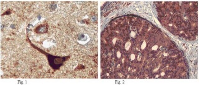
Immunohistochemistry Analysis: A representative lot detected Rootletin in Immunohistochemistry applications (Yang, J., et. al. (2002). J Cell Biol. 159(3):431-40; Sun, X., etl al. (2016). Proc Natl Acad Sci USA. 113(21):E2925-34; Yadav, S.P., et. al. (2016). Development. 143(9):1491-501; Pawlyk, B.S., et. al. (2016). Gene Ther. 23(2):196-204; Sun X., et. al. (2012). Cilia. 1(1):21; Liu, X., et. al. (2007). Proc Antl Acad Sci USA. 104(11):4413-8; Bahe, S., et. al. (2005). J Cell Biol. 171(1):27-33; Yang, J., et. al. (2005). Mol Cell Biol. 25(10):4129-37; Hong, D.H, et. al. (2003). Invest Ophthalmol Vis Sci. 44(6):2413-21).

Quality

Evaluated by Western Blotting in mouse retina tissue lysate.

Western Blotting Analysis: A 1,000 dilution of this antibody detected Rootletin in 10 µg of mouse retina tissue lysate.

Target description

~227 kDa observed; 226.95 kDa calculated. Uncharacterized bands may be observed in some lysate(s).

Physical form

Affinity Purified
Purified chicken polyclonal antibody in PBS with 50% glycerol and without azide.

Storage and Stability

Stable for 1 year at -20°C from date of receipt.

Handling Recommendations: Upon receipt and prior to removing the cap, centrifuge the vial and gently mix the solution. Aliquot into microcentrifuge tubes and store at -20°C. Avoid repeated freeze/thaw cycles, which may damage IgG and affect product performance.

Other Notes

Concentration: Please refer to lot specific datasheet.

Disclaimer

Unless otherwise stated in our catalog or other company documentation accompanying the product(s), our products are intended for research use only and are not to be used for any other purpose, which includes but is not limited to, unauthorized commercial uses, in vitro diagnostic uses, ex vivo or in vivo therapeutic uses or any type of consumption or application to humans or animals.

Not finding the right product?  

Try our Product Selector Tool.

Storage Class Code

10 - Combustible liquids

WGK

WGK 2


Certificates of Analysis (COA)

Search for Certificates of Analysis (COA) by entering the products Lot/Batch Number. Lot and Batch Numbers can be found on a product’s label following the words ‘Lot’ or ‘Batch’.

Already Own This Product?

Find documentation for the products that you have recently purchased in the Document Library.

Visit the Document Library

Rachel E Turn et al.
Molecular biology of the cell, 32(8), 800-822 (2021-02-18)
ELMOD2 is a GTPase-activating protein with uniquely broad specificity for ARF family GTPases. We previously showed that it acts with ARL2 in mitochondrial fusion and microtubule stability and with ARF6 during cytokinesis. Mouse embryonic fibroblasts deleted for ELMOD2 also displayed
Ying Hsu et al.
PLoS genetics, 13(10), e1007057-e1007057 (2017-10-20)
Genetic mutations disrupting the structure and function of primary cilia cause various inherited retinal diseases in humans. Bardet-Biedl syndrome (BBS) is a genetically heterogeneous, pleiotropic ciliopathy characterized by retinal degeneration, obesity, postaxial polydactyly, intellectual disability, and genital and renal abnormalities.
Skylar I Dewees et al.
Molecular biology of the cell, 33(4), ar33-ar33 (2022-02-24)
The ARF family of regulatory GTPases is ancient, with 16 members predicted to have been present in the last eukaryotic common ancestor. Our phylogenetic profiling of paralogues in diverse species identified four family members whose presence correlates with that of
Kristen N Haggerty et al.
PLoS biology, 22(1), e3002467-e3002467 (2024-01-08)
Photoreceptor cells in the vertebrate retina have a highly compartmentalized morphology for efficient phototransduction and vision. Rhodopsin, the visual pigment in rod photoreceptors, is densely packaged into the rod outer segment sensory cilium and continuously renewed through essential synthesis and
Rachel E Turn et al.
Molecular biology of the cell, mbcE21090443-mbcE21090443 (2021-11-25)
ELMODs are a family of three mammalian paralogs that display GTPase activating protein (GAP) activity towards a uniquely broad array of ADP-ribosylation factor (ARF) family GTPases that includes ARF-like (ARL) proteins. ELMODs are ubiquitously expressed in mammalian tissues, highly conserved

Our team of scientists has experience in all areas of research including Life Science, Material Science, Chemical Synthesis, Chromatography, Analytical and many others.

Contact Technical Service