Skip to Content
Merck
All Photos(1)

Key Documents

108M-9

Sigma-Aldrich

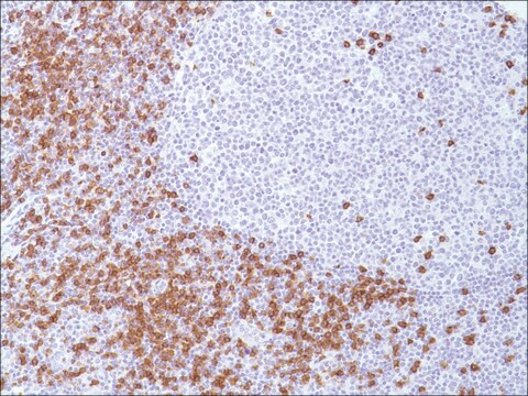
CD8 (C8/144B) Mouse Monoclonal Antibody

Sign Into View Organizational & Contract Pricing


About This Item

UNSPSC Code:
12352203
NACRES:
NA.41
clone:
C8/144B, monoclonal
application:
IHC (p)
species reactivity:
human
technique(s):
immunohistochemistry (formalin-fixed, paraffin-embedded sections): 1:25-1:100
citations:
6

biological source

mouse

Quality Level

100
500

conjugate

unconjugated

antibody form

diluted ascites fluid

antibody product type

primary antibodies

clone

C8/144B, monoclonal

description

For In Vitro Diagnostic Use in Select Regions (See Chart)

form

buffered aqueous solution

species reactivity

human

packaging

vial of 0.1 mL concentrate (108M-94)
vial of 0.5 mL concentrate (108M-95)
bottle of 1.0 mL predilute (108M-97)
vial of 1.0 mL concentrate (108M-96)
bottle of 7.0 mL predilute (108M-98)

manufacturer/tradename

Cell Marque

technique(s)

immunohistochemistry (formalin-fixed, paraffin-embedded sections): 1:25-1:100

isotype

IgG1κ

control

tonsil

shipped in

wet ice

storage temp.

2-8°C

visualization

membranous

Gene Information

human ... CD8A(925)

General description

Anti-CD8 is a T cell marker for the detection of cytotoxic/suppresser cells of blood lymphocytes. CD8 is also detected on NK cells, most thymocytes, a subpopulation of null cells and bone marrow cells. This antibody, along with other markers, can be used to distinguish between reactive and neoplastic T-cells.

Quality


IVD

IVD

IVD

RUO

Linkage

CD8 Positive Control Slides, Product No. 108S, are available for immunohistochemistry (formalin-fixed, paraffin-embedded sections).

Physical form

Solution in Tris Buffer, pH 7.3-7.7, with 1% BSA and <0.1% Sodium Azide

Preparation Note

Download the IFU specific to your product lot and formatNote: This requires a keycode which can be found on your packaging or product label.

Other Notes

For Technical Service please contact: 800-665-7284 or email: service@cellmarque.com

Legal Information

Cell Marque is a trademark of Merck KGaA, Darmstadt, Germany

Not finding the right product?  

Try our Product Selector Tool.


Choose from one of the most recent versions:

Certificates of Analysis (COA)

Lot/Batch Number

Don't see the Right Version?

If you require a particular version, you can look up a specific certificate by the Lot or Batch number.

Already Own This Product?

Find documentation for the products that you have recently purchased in the Document Library.

Visit the Document Library

M L Rossi et al.
Journal of clinical pathology, 41(3), 314-319 (1988-03-01)
Twenty four meningiomas (17 benign and seven "atypical" were reacted with a panel of monoclonal antibodies to macrophages, lymphocytes, and HLA DR antigens. All the tumours contained macrophages but these cells were more numerous in the atypical meningiomas. Lymphocytes, almost
D Y Mason et al.
Journal of clinical pathology, 45(12), 1084-1088 (1992-12-01)
To evaluate whether cytotoxic/suppressor T cells can be detected in paraffin wax embedded human tissue samples using antibodies to a synthetic CD8 peptide sequence. Polyclonal and monoclonal antibodies were raised against a 13 amino acid peptide sequence from the cytoplasmic
Immunohistological analysis of human lymphoma: correlation of histological and immunological categories.
H Stein et al.
Advances in cancer research, 42, 67-147 (1984-01-01)
F Phan-Dinh-Tuy et al.
Molecular immunology, 19(12), 1649-1654 (1982-12-01)
Three human lymphocyte differentiation antigens, specific of the entire T-cell population, of the helper/inducer T-cell subset, and of the cytotoxic/suppressor T-cell subset have been identified, using mouse monoclonal antibodies obtained from Dr. P. Kung. Various T-cell populations were radio-labelled, the
J D Nuckols et al.
Journal of cutaneous pathology, 26(4), 169-175 (1999-05-21)
Differentiation between mycosis fungoides (MF) and cutaneous inflammatory processes can usually be made on clinical and histologic grounds. In difficult cases, immunohistochemical studies can be helpful since MF infiltrates usually contain a predominance of CD4+ lymphocytes, while most inflammatory lesions

Our team of scientists has experience in all areas of research including Life Science, Material Science, Chemical Synthesis, Chromatography, Analytical and many others.

Contact Technical Service