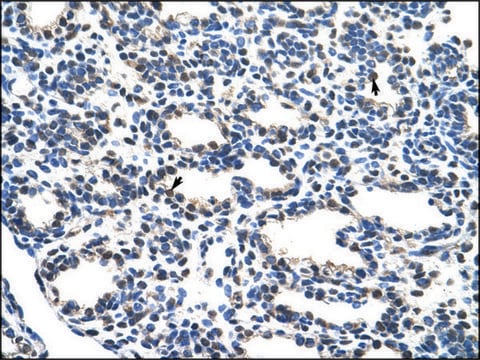
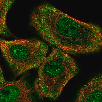
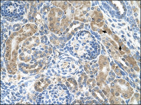
DCP1A is an mRNA decapping enzyme that is involved in TGF-β signaling. DCP1A is known to be hyper-phosphorylated during mitosis, cellular stress, neuronal differentiation and brain development. DCP1A has also been implicated in translational arrest. Rabbit Anti-DCP1A antibody recognizes rat, human, chicken, bovine, canine, zebrafish, and mouse DCP1A.
Immunogen
Synthetic peptide directed towards the N terminal region of human DCP1A
Application
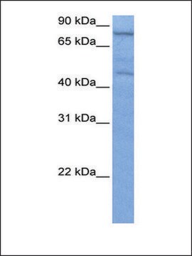
Rabbit Anti-DCP1A antibody is suitable for western blot applications at a concentration of 1.25 μg/ml.
Biochem/physiol Actions
Decapping is a key step in general and regulated mRNA decay. The protein encoded by this gene is a decapping enzyme. This protein and another decapping enzyme form a decapping complex, which interacts with the nonsense-mediated decay factor hUpf1 and may be recruited to mRNAs containing premature termination codons. This protein also participates in the TGF-beta signaling pathway.Decapping is a key step in general and regulated mRNA decay. The protein encoded by this gene is a decapping enzyme. This protein and another decapping enzyme form a decapping complex, which interacts with the nonsense-mediated decay factor hUpf1 and may be recruited to mRNAs containing premature termination codons. This protein also participates in the TGF-beta signaling pathway.
Sequence
Synthetic peptide located within the following region: MEALSRAGQEMSLAALKQHDPYITSIADLTGQVALYTFCPKANQWEKTDI
Physical form
Purified antibody supplied in 1x PBS buffer with 0.09% (w/v) sodium azide and 2% sucrose.
Disclaimer
Unless otherwise stated in our catalog or other company documentation accompanying the product(s), our products are intended for research use only and are not to be used for any other purpose, which includes but is not limited to, unauthorized commercial uses, in vitro diagnostic uses, ex vivo or in vivo therapeutic uses or any type of consumption or application to humans or animals.
Decapping protein 1a (Dcp1a) is found in P-bodies and functions in mRNA cap removal prior to its degradation. The function and binding partners of Dcp1a have been thoroughly studied, however its expression pattern is still unclear. In this study we
The Journal of biological chemistry, 289(7), 3936-3949 (2014-01-03)
We have shown previously that poliovirus infection disrupts cytoplasmic P-bodies in infected mammalian cells. During the infectious cycle, poliovirus causes the directed cleavage of Dcp1a and Pan3, coincident with the dispersion of P-bodies. We now show that expression of Dcp1a
Processing bodies (PBs) are non-membranous cytoplasmic structures found in all eukaryotes. Many of their components such as the Dcp1 and Dcp2 proteins are highly conserved. Using live-cell imaging we found that PB structures disassembled as cells prepared for cell division
Questions
Reviews
★★★★★ No rating value
Active Filters
Our team of scientists has experience in all areas of research including Life Science, Material Science, Chemical Synthesis, Chromatography, Analytical and many others.