Skip to Content
Merck
All Photos(2)

Documents

T1948

Sigma-Aldrich

Anti-TRF1 antibody, Mouse monoclonal

~2 mg/mL, clone TRF-78, purified from hybridoma cell culture

Synonym(s):

Anti-Telomeric Repeat Binding Factor 1

Sign Into View Organizational & Contract Pricing


About This Item

MDL number:
UNSPSC Code:
12352203
NACRES:
NA.41

biological source

mouse

Quality Level

conjugate

unconjugated

antibody form

purified from hybridoma cell culture

antibody product type

primary antibodies

clone

TRF-78, monoclonal

form

buffered aqueous solution

mol wt

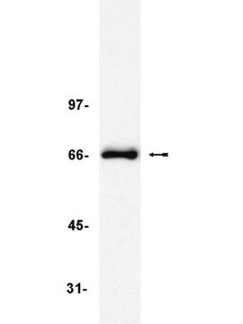
antigen ~70 kDa by SDS-PAGE

species reactivity

human

packaging

antibody small pack of 25 μL

concentration

~2 mg/mL

technique(s)

indirect ELISA: suitable
microarray: suitable
western blot: 2-4 μg/mL using HeLa nuclear extract

isotype

IgG1

UniProt accession no.

shipped in

dry ice

storage temp.

−20°C

target post-translational modification

unmodified

Gene Information

human ... TERF1(7013)

General description

Monoclonal Anti-TRF1 (mouse IgG1 isotype) is derived from the TRF-78 hybridoma produced by the fusion of mouse myeloma cells and splenocytes from mice immunized with a human TRF1 protein produced in baculovirus. TRF1 and TRF2 (TTAGGG repeat binding factors) are two major proteins that bind to human telomers. TRF1 has a DNA binding domain with high homology to the Myb family of transcription factors. Unlike the Myb family that contains only one DNA binding motif, TRF1 has multiple of this motif.

Specificity

Monoclonal Anti-TRF1 recognizes human TRF1.

Immunogen

human TRF1 protein produced in baculovirus.

Application

Monoclonal Anti-TRF1 antibody produced in mouse has been used in Western blotting and enzyme linked immunosorbent assay (ELISA).

Biochem/physiol Actions

TRF1 (TTAGGG repeat binding factor 1) has a negative effect on the length of the telomer. Overexpression of TRF1 in cancer cells that contain telomerase activity, causes the shortening of the length of their telomers. While inhibition of TRF1 causes the elongation of telomers. It was shown that the level of TRF1 in the cells does not affect the expression of the telomerase protein. This suggests that TRF1 may act directly on the activity of the telomerase protein. Tankyrase is a protein that interacts with TRF1 and its C-terminal region is homologous to poly (adenosine dinucleotide phosphate (ADP)-ribose) polymerase (PARP). In response to DNA damage, the PARP protein mediates ADP-ribose polymers of protein acceptors. In vitro studies have shown that tankyrase is responsible for that polyribosylation of TRF1, which in turn abolishes its ability to bind telomers.

Physical form

Solution in 0.01 M phosphate buffered saline, pH 7.4, and 15 mM sodium azide.

Disclaimer

Unless otherwise stated in our catalog or other company documentation accompanying the product(s), our products are intended for research use only and are not to be used for any other purpose, which includes but is not limited to, unauthorized commercial uses, in vitro diagnostic uses, ex vivo or in vivo therapeutic uses or any type of consumption or application to humans or animals.

Not finding the right product?  

Try our Product Selector Tool.

Storage Class Code

10 - Combustible liquids

WGK

WGK 2


Certificates of Analysis (COA)

Search for Certificates of Analysis (COA) by entering the products Lot/Batch Number. Lot and Batch Numbers can be found on a product’s label following the words ‘Lot’ or ‘Batch’.

Already Own This Product?

Find documentation for the products that you have recently purchased in the Document Library.

Visit the Document Library

Xinyu Ci et al.
Oncotarget, 6(35), 38079-38092 (2015-10-17)
Bortezomib inhibits the ubiquitin/proteasome pathway to achieve its anti-cancer effect and its well characterized activity is the NF-κB inhibition through which the anti-apoptotic bcl-2 expression is down-regulated and apoptosis is subsequently induced. However, the downstream molecular targets of bortezomib are
Bortezomib-mediated down-regulation of telomerase and disruption of telomere homeostasis contributes to apoptosis of malignant cells
Ci X, et al.
Oncotarget, 6(35), 38079-38079 (2015)
Juan Ge et al.
Aging, 11(7), 1965-1976 (2019-03-31)
It has been well recognized that oocyte quality declines in aging animals. However, to date, the underlying mechanism remains to be explored. In the present study, we report that oocytes and embryos from aged mice (42-45 weeks old) display the
Protection of mammalian telomeres
de Lange T
Oncogene, 21(4), 532-532 (2002)
Man Zhang et al.
Aging, 11(12), 3919-3938 (2019-06-18)
Mitochondria change their shape through fusion and fission in order to adapt to their metabolic milieu. Mitofusin-2 (MFN2) is a key regulatory protein in this process, mediating mitochondrial fusion and interaction with endoplasmic reticulum. Targeted deletion of Mfn2 in oocytes

Our team of scientists has experience in all areas of research including Life Science, Material Science, Chemical Synthesis, Chromatography, Analytical and many others.

Contact Technical Service