Skip to Content
Merck
All Photos(2)

Key Documents

MABE1785

Sigma-Aldrich

Anti-Ubiquitinated Histone H2B (Lys123) Antibody, clone 1B3F12/A9

clone 1B3F12/A9, from mouse

Synonym(s):

Histone H2B K123 Ub1, Histone H2B.2 UbK123

Sign Into View Organizational & Contract Pricing


About This Item

UNSPSC Code:
12352203
eCl@ss:
32160702
NACRES:
NA.41

biological source

mouse

Quality Level

antibody form

purified immunoglobulin

antibody product type

primary antibodies

clone

1B3F12/A9, monoclonal

species reactivity

yeast, human

packaging

antibody small pack of 25 μg

technique(s)

ChIP: suitable
western blot: suitable

isotype

IgG1κ

NCBI accession no.

UniProt accession no.

shipped in

ambient

target post-translational modification

unmodified

Gene Information

Saccharomyces cerevisiae ... Htb2(852284)
human ... H2BC1(255626)

General description

Histone H2B.2 (UniProt: P02294) is encoded by the HTB2 (also known as H2B2) gene (Gene ID: 852284) in Saccharomyces cerevisiae (yeast). Histones are basic nuclear proteins that are responsible for the nucleosome structure of chromatin in eukaryotes. Two molecules of each of the four core histones (H2A, H2B, H3, and H4) form an octamer, around which DNA is wrapped in repeating units, called nucleosomes, which limits DNA accessibility to the cellular machineries, which require DNA as a template. Histones thereby play a central role in transcription regulation, DNA repair, DNA replication and chromosomal stability. DNA accessibility is regulated via a complex set of post-translational modifications of histones, also called histone code, and nucleosome remodeling. Histone H2B is one of the main histone proteins involved in the structure of chromatin in eukaryotic cells. Featuring a main globular domain and a long N terminal tail, H2B is involved with the structure of the nucleosomes of the ′beads on a string′ structure. Histone H2B can be monoubiquitinated on Lysine123 by RAD6/UBC2-BRE1 complex to form H2BK123Ub1 complex that gives a specific tag for epigenetic transcriptional activation and is also a prerequisite for H3K4me and H3K79me formation. H2BK123ub1 is reported to modulates the formation of double-strand breaks during meiosis and is a required for DNA-damage checkpoint activation. Histone H2B is phosphorylated by STE20 to form H2BS10ph during progression through meiotic prophase and may be correlated with chromosome condensation. Histone H2B can undergo acetylation by GCN5, a component of the SAGA complex, to form H2BK11ac and H2BK16ac. Acetylation of N-terminal lysines and particularly formation of H2BK11acK16ac has a positive effect on transcription.

Specificity

Clone 1B3F12/A9 detects K123 ubiquitinated histone H2B in human and yeast. It targets an epitope within 16 amino acids from the C-terminal region of ubiquitinated yeast Histone H2B.

Immunogen

Conjugated linear peptide corresponding to 16 amino acids from the C-terminal region of yeast Histone H2B ubiquitinated at Lysine 123.

Application

Anti-Ubiquitinated Histone H2B (Lys123), clone 1B3F12/A9 Antibody, Cat. No. MABE1785, is a highly specific mouse monoclonal antibody, that targets ubiquitinated Histone H2B and has been tested in Chromatin Immunoprecipitation (ChIP) and Western Blotting.
Research Category
Epigenetics & Nuclear Function
Western Blotting Analysis: 2 µg/mL from a representative lot detected Ubiquitinated Histone H2B (Lys123) in wild type whole cell yeast extracts and ubp8 knockout whole cell yeast extracts.

Chromatin Immunoprecipitation (ChIP) Analysis: A representative lot detected Ubiquitinated Histone H2B (Lys123) in H2BK123ub relative to H2B at the coding sequence of the SPA2 gene in budding yeast (relative to WT) (Courtesy of Fred van Leeuwen, Ph.D., Division of Gene Regulation B4, The Netherlands Cancer Institute Plesmanlaan 121, Amsterdam Netherlands).

Quality

Evaluated by Western Blotting in HeLa cell acid extract.

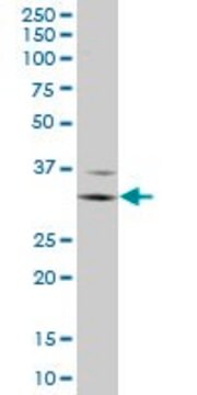
Western Blotting Analysis: 2 µg/mL of this antibody detected Ubiquitinated Histone H2B (Lys123) in HeLa cell acid extract.

Target description

~23 kDa observed; 14.25 kDa calculated. Uncharacterized bands may be observed in some lysate(s).

Physical form

Format: Purified
Protein G purified
Purified mouse monoclonal antibody IgG1 in buffer containing 0.1 M Tris-Glycine (pH 7.4), 150 mM NaCl with 0.05% sodium azide.

Storage and Stability

Stable for 1 year at 2-8°C from date of receipt.

Other Notes

Concentration: Please refer to lot specific datasheet.

Disclaimer

Unless otherwise stated in our catalog or other company documentation accompanying the product(s), our products are intended for research use only and are not to be used for any other purpose, which includes but is not limited to, unauthorized commercial uses, in vitro diagnostic uses, ex vivo or in vivo therapeutic uses or any type of consumption or application to humans or animals.

Not finding the right product?  

Try our Product Selector Tool.

Storage Class Code

12 - Non Combustible Liquids

WGK

WGK 1

Flash Point(F)

does not flash

Flash Point(C)

does not flash


Certificates of Analysis (COA)

Search for Certificates of Analysis (COA) by entering the products Lot/Batch Number. Lot and Batch Numbers can be found on a product’s label following the words ‘Lot’ or ‘Batch’.

Already Own This Product?

Find documentation for the products that you have recently purchased in the Document Library.

Visit the Document Library

Our team of scientists has experience in all areas of research including Life Science, Material Science, Chemical Synthesis, Chromatography, Analytical and many others.

Contact Technical Service