Skip to Content
Merck
All Photos(1)

Documents

AQ303F

Sigma-Aldrich

Goat Anti-Mouse IgG Antibody, F(ab′)2, FITC conjugate

1 mg/mL, Chemicon®

Synonym(s):

Goat anti-Mouse IgG FITC

Sign Into View Organizational & Contract Pricing


About This Item

UNSPSC Code:
12352203
eCl@ss:
32160702
NACRES:
NA.46

biological source

goat

Quality Level

conjugate

FITC conjugate

antibody form

F(ab′)2 fragment of affinity isolated antibody

antibody product type

secondary antibodies

clone

polyclonal

species reactivity

mouse

manufacturer/tradename

Chemicon®

concentration

1 mg/mL

technique(s)

immunofluorescence: suitable

shipped in

wet ice

target post-translational modification

unmodified

General description

Immunoglobulin G (IgG), is one of the most abundant proteins in human serum with normal levels between 8-17 mg/mL in adult blood. IgG is important for our defence against microorganisms and the molecules are produced by B lymphocytes as a part of our adaptive immune response. The IgG molecule has two separate functions; to bind to the pathogen that elicited the response and to recruit other cells and molecules to destroy the antigen. The variability of the IgG pool is generated by somatic recombination and the number of specificities in an individual at a given time point is estimated to be 1011 variants.
The reagent is an anti-mouse antibody F(ab′)2 fragment from goat. The F(ab′)2 fragments are obtained by pepsin cleavage. The fragments are conjugated to fluorescein isothiocyanate and stabilized in buffer. The molar ratio of fluorescein: protein is: 3 - 7.

PURIFICATION: The goat-lgG was purified by affinity chromatography and absorbed to remove cross-reactivity to human immunoglobulins.

Specificity

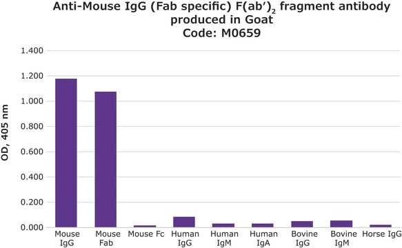
Specific for mouse IgG, heavy and light chain. The cross-reactivities of the anti-mouse IgG antibody were tested in an ELISA. Minimum cross-reactivity to human IgG.

Application

Immunohistochemistry: 1:200-1:500 (Coligan et al. 1997; Harlow & Lane 1988; Bullock & Petrusz 1982; Javois 1994)

Immunocytochemistry: 1:200-1:500 (Coligan et al. 1997; Harlow & Lane 1988; Bullock & Petrusz 1982; Javois 1994)

Flow cytometery: 1 μg per 1 x 10E6 cells (Coligan et al. 1997; Harlow & Lane 1988; Javois 1994) FITC absorption peak is 488-492nm, its emission peak is 520nm.

Optimal working dilutions must be determined by the end user.
Research Category
Secondary & Control Antibodies
Research Sub Category
Fragment Specific Secondary Antibodies
This Goat anti-Mouse IgG Antibody, F(ab′)2, FITC conjugate is validated for use in IF for the detection of Mouse IgG.

Physical form

ImmunoAffinity Purified
Liquid in 0.02M Phosphate Buffer. 0.25 M NaCl, pH 7.6 with 15 mg/mL BSA and 0.01% Sodium azide.

Storage and Stability

The antibody conjugate solution is stable at 2–8°C for 12 months. Do not store in a diluted format.

Legal Information

CHEMICON is a registered trademark of Merck KGaA, Darmstadt, Germany

Disclaimer

Unless otherwise stated in our catalog or other company documentation accompanying the product(s), our products are intended for research use only and are not to be used for any other purpose, which includes but is not limited to, unauthorized commercial uses, in vitro diagnostic uses, ex vivo or in vivo therapeutic uses or any type of consumption or application to humans or animals.

Not finding the right product?  

Try our Product Selector Tool.

Storage Class Code

12 - Non Combustible Liquids

WGK

WGK 2

Flash Point(F)

Not applicable

Flash Point(C)

Not applicable


Certificates of Analysis (COA)

Search for Certificates of Analysis (COA) by entering the products Lot/Batch Number. Lot and Batch Numbers can be found on a product’s label following the words ‘Lot’ or ‘Batch’.

Already Own This Product?

Find documentation for the products that you have recently purchased in the Document Library.

Visit the Document Library

Lijun Xin et al.
Aging cell, 14(6), 1122-1126 (2015-08-08)
Aging confers increased susceptibility to common pathogens including influenza A virus. Despite shared vulnerability to infection with advancing age in humans and rodents, the relatively long time required for immune senescence to take hold practically restricts the use of naturally
Daniel Sobrido-Cameán et al.
The Journal of comparative neurology, 531(1), 58-88 (2022-09-24)
The expression of the corticotropin-releasing hormone (PmCRH) and the CRH-binding protein (PmCRHBP) mRNAs was studied by in situ hybridization in the brain of prolarvae, larvae, and adults of the sea lamprey Petromyzon marinus. We also generated an antibody against the
Antón Barreiro-Iglesias et al.
The Journal of comparative neurology, 525(17), 3683-3704 (2017-08-05)
We employed an anti-transducin antibody (Gαt-S), in combination with other markers, to characterize the Gαt-S-immunoreactive (ir) system in the CNS of the sea lamprey, Petromyzon marinus. Gαt-S immunoreactivity was observed in some neuronal populations and numerous fibers distributed throughout the
Single-walled carbon nanotubes increase pandemic influenza A H1N1 virus infectivity of lung epithelial cells.
Sanpui, P; Zheng, X; Loeb, JC; Bisesi, JH; Khan, IA; Afrooz, AR; Liu, K; Badireddy et al.
Particle and Fibre Toxicology null
Ramón Anadón et al.
The Journal of comparative neurology, 532(2), e25590-e25590 (2024-02-09)
Gamma-aminobutyric acid (GABA) is the main inhibitory neurotransmitter in the central nervous system (CNS) of vertebrates. Immunohistochemical techniques with specific antibodies against GABA or against its synthesizing enzyme, glutamic acid decarboxylase (GAD) allowed characterizing GABAergic neurons and fibers in the

Our team of scientists has experience in all areas of research including Life Science, Material Science, Chemical Synthesis, Chromatography, Analytical and many others.

Contact Technical Service