06-896
Anti-GFP (Green Fluorescent Protein) Antibody
Upstate®, from chicken
Synonym(s):
Green fluorescent protein
Sign Into View Organizational & Contract Pricing
All Photos(1)
About This Item
UNSPSC Code:
12352203
eCl@ss:
32160702
NACRES:
NA.41
Recommended Products
biological source
chicken
Quality Level
antibody form
purified immunoglobulin
antibody product type
primary antibodies
clone
polyclonal
species reactivity
vertebrates
manufacturer/tradename
Upstate®
technique(s)
immunocytochemistry: suitable
western blot: suitable
isotype
IgY
UniProt accession no.
shipped in
dry ice
target post-translational modification
unmodified
General description
Green fluorescent protein (GFP) is a 29 kDa fluorescent protein isolated from the jellyfish Aequorea victoria. Upon exposure to blue light (488 nm), GFP strongly fluoresces (Emission max: 507 nm). GFP can be fused to proteins without significantly interfering with their assembly and function, making GFP a powerful tool for monitoring gene expression and protein localization in living cells.
Specificity
Recognizes green fluorescent protein (GFP), Mr 30 kDa, and GFP-fusion proteins.
Immunogen
His-tagged green fluorescent protein of Aequorea victoria.
Application
Anti-GFP (Green Fluorescent Protein) Antibody detects level of GFP (Green Fluorescent Protein) & has been published & validated for use in IC & WB.
Immunocytochemistry:
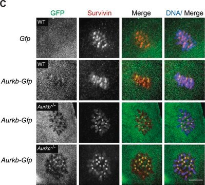
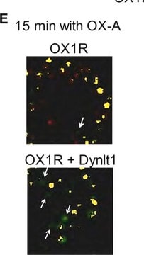
This antibody was reported to show positive immunostaining in cells transfected with an expression vector encoding Green Fluorescent Protein.
This antibody was reported to show positive immunostaining in cells transfected with an expression vector encoding Green Fluorescent Protein.
Research Category
Epitope Tags & General Use
Epitope Tags & General Use
Research Sub Category
Epitope Tags
Epitope Tags
Quality
Routinely evaluated by Western Blot on rat brain lysate containing purified GFP (Catalog #14-392) or a GFP-fusion protein from a transfected E. coli cell lysate.
Western Blot Analysis:
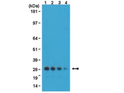
0.5-2 μg/mL of this lot detected Green Fluorescent Protein from 50 μg of rat brain lysate containing 25 ng of purified Green Fluorescent Protein (Catalog # 14-392). A previous lot of this antibody also detected a GFP-fusion protein from a transfected E. coli cell lysate.
Western Blot Analysis:
0.5-2 μg/mL of this lot detected Green Fluorescent Protein from 50 μg of rat brain lysate containing 25 ng of purified Green Fluorescent Protein (Catalog # 14-392). A previous lot of this antibody also detected a GFP-fusion protein from a transfected E. coli cell lysate.
Target description
30 kDa
Physical form
Ammonium Sulfate Precipitation and PEG
Format: Purified
Purified chicken polyclonal IgY from egg yolk in buffer containing 70% storage buffer (0.1M Tris-glycine, pH 7.4, 0.15M NaCl, 0.05% sodium azide) and 30% glycerol. Store at -20°C.
Storage and Stability
Stable for 1 year at -20°C from date of receipt.
Handling Recommendations: Upon receipt, and prior to removing the cap, centrifuge the vial and gently mix the solution. Aliquot into microcentrifuge tubes and store at -20°C. Avoid repeated freeze/thaw cycles, which may damage IgG and affect product performance. Note: Variability in freezer temperatures below -20°C may cause glycerolcontaining solutions to become frozen during storage.
Handling Recommendations: Upon receipt, and prior to removing the cap, centrifuge the vial and gently mix the solution. Aliquot into microcentrifuge tubes and store at -20°C. Avoid repeated freeze/thaw cycles, which may damage IgG and affect product performance. Note: Variability in freezer temperatures below -20°C may cause glycerolcontaining solutions to become frozen during storage.
Analysis Note
Control
Rat brain lysate, cells expressing GFP protein.
Rat brain lysate, cells expressing GFP protein.
Other Notes
Concentration: Please refer to the Certificate of Analysis for the lot-specific concentration.
Legal Information
UPSTATE is a registered trademark of Merck KGaA, Darmstadt, Germany
Disclaimer
Unless otherwise stated in our catalog or other company documentation accompanying the product(s), our products are intended for research use only and are not to be used for any other purpose, which includes but is not limited to, unauthorized commercial uses, in vitro diagnostic uses, ex vivo or in vivo therapeutic uses or any type of consumption or application to humans or animals.
Not finding the right product?
Try our Product Selector Tool.
recommended
Storage Class Code
10 - Combustible liquids
WGK
WGK 1
Certificates of Analysis (COA)
Search for Certificates of Analysis (COA) by entering the products Lot/Batch Number. Lot and Batch Numbers can be found on a product’s label following the words ‘Lot’ or ‘Batch’.
Already Own This Product?
Find documentation for the products that you have recently purchased in the Document Library.
Customers Also Viewed
Green fluorescent protein: applications in cell biology.
Gerdes, H H and Kaether, C
Febs Letters, 389, 44-47 (1996)
Nicolás Pírez et al.
Biology open, 8(1) (2018-12-12)
In the fruit fly, Drosophila melanogaster, the daily cycle of rest and activity is a rhythmic behavior that relies on the activity of a small number of neurons. The small ventral lateral neurons (sLNvs) are considered key in the control
Corette J Wierenga et al.
PloS one, 5(12), e15915-e15915 (2011-01-07)
The use of transgenic mice in which subtypes of neurons are labeled with a fluorescent protein has greatly facilitated modern neuroscience research. GAD65-GFP mice, which have GABAergic interneurons labeled with GFP, are widely used in many research laboratories, although the
MARK2/Par-1 guides the directionality of neuroblasts migrating to the olfactory bulb.
Sheyla Mejia-Gervacio,Kerren Murray,Tamar Sapir,Richard Belvindrah,Orly Reiner,Pierre-Marie Lledo
Molecular and Cellular Neurosciences null
Richard Mark White et al.
Cell stem cell, 2(2), 183-189 (2008-03-29)
The zebrafish is a useful model for understanding normal and cancer stem cells, but analysis has been limited to embryogenesis due to the opacity of the adult fish. To address this, we have created a transparent adult zebrafish in which
Our team of scientists has experience in all areas of research including Life Science, Material Science, Chemical Synthesis, Chromatography, Analytical and many others.
Contact Technical Service